Ravichandran Rajkumar1,2,3, Gereon Johannes Schnellbächer2, Hasan Sbaihat1,2, N. Jon Shah1,4,5,6, Tanja Veselinović2, and Irene Neuner1,2,4
1Institute of Neuroscience and Medicine - 4 (Medical Imaging Physics), Forschungszentrum Juelich GmbH, Jülich, Germany, 2Department of Psychiatry, Psychotherapy and Psychosomatics, RWTH Aachen University, Aachen, Germany, 34JARA – BRAIN – Translational Medicine, Aachen, Germany, 4JARA – BRAIN – Translational Medicine, Aachen, Germany, 5Department of Neurology, RWTH Aachen University, Aachen, Germany, 6Institute of Neuroscience and Medicine, INM-11, Forschungszentrum Jülich GmbH, Jülich, Germany
1Institute of Neuroscience and Medicine - 4 (Medical Imaging Physics), Forschungszentrum Juelich GmbH, Jülich, Germany, 2Department of Psychiatry, Psychotherapy and Psychosomatics, RWTH Aachen University, Aachen, Germany, 34JARA – BRAIN – Translational Medicine, Aachen, Germany, 4JARA – BRAIN – Translational Medicine, Aachen, Germany, 5Department of Neurology, RWTH Aachen University, Aachen, Germany, 6Institute of Neuroscience and Medicine, INM-11, Forschungszentrum Jülich GmbH, Jülich, Germany
This 7T fMRI pilot study
suggests that changes in functional parameters within the ECN may be due to
executive function impairments in depressed patients
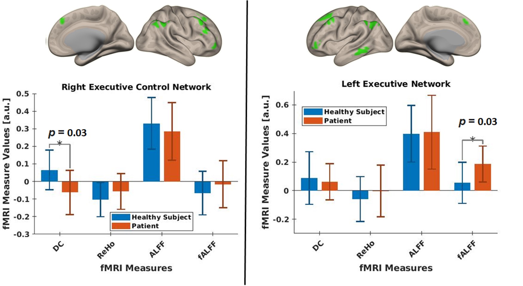
Fig. 2: Bar chart with the standard
deviation for each RS-fMRI measure within the right executive control network
(RECN, right side) and the left executive control network (LECN, left side)
between a healthy subject and a depressed patient. Right and left view of the
masks of the RECN and LECN used in the analysis are shown above the
corresponding bar chart.
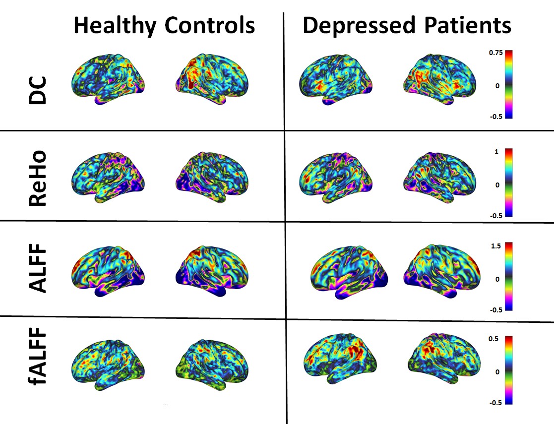
Fig. 1: Mean RS-fMRI measures of
healthy subjects (right column) and depressed patients (left column). The fMRI
measures degree centrality (DC, top row), regional homogeneity (ReHo, second
row), amplitude of low frequency fluctuations (ALFF, third row) and fractional
ALFF (bottom row) are shown in right, and left views.