Katherine E Lawrence1, Leila Nabulsi1, Vigneshwaran Santhalingam1, Zvart Abaryan1, Julio E Villalon-Reina1, Talia M Nir1, Iyad Ba Gari1, Alyssa H Zhu1, Elizabeth Haddad1, Alexandra M Muir1, Neda Jahanshad1, and Paul M Thompson1
1University of Southern California, Marina del Rey, CA, United States
1University of Southern California, Marina del Rey, CA, United States
Using traditional (DTI) and
advanced (TDF, NODDI, MAPMRI) diffusion-weighted MRI models, we found that
advanced diffusion approaches exhibited the greatest sensitivity to age and sex
effects on white matter microstructure.

Figure 3. Normative
centile reference curves for each diffusion-weighted MRI metric, computed using
quantile regression for single-shell metrics in (A) males and (B) females, and
multi-shell metrics in (C) males and (D) females. Solid colored lines, ordered
from lightest to darkest, indicate the following centiles: 5th, 25th,
50th, 75th, 95th; blue lines indicate male
participants, and red lines indicate female participants. Gray overlay reflects
kernel density (darker=greater data overlap).
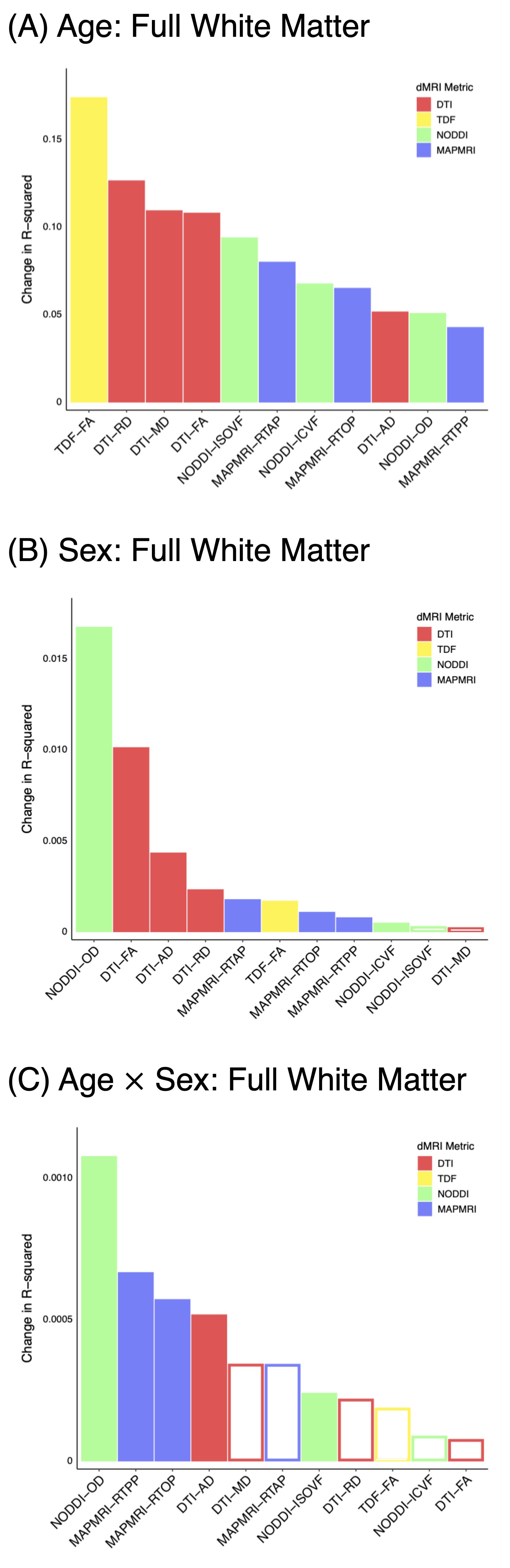
Figure 1. Effect of
age (A), participant sex (B), and their interaction (C) on whole-skeleton white
matter microstructure when modeling age as a continuous variable using
fractional polynomials. Filled bars indicate a significant association
(uncorrected), whereas hollow bars indicate the association did not attain
statistical significance. Results were essentially identical after FDR
correction for the number of metrics, except the age by sex interaction no
longer attained significance for NODDI-ISOVF.