Marina Manso Jimeno1,2, Keerthi Sravan Ravi2,3, John Thomas Vaughan Jr.2,3, Dotun Oyekunle4, Godwin Ogbole4, and Sairam Geethanath2
1Biomedical Engineering, Columbia University, New York, NY, United States, 2Columbia Magnetic Resonance Research Center (CMRRC), New York, NY, United States, 3Columbia University, New York, NY, United States, 4Radiology, University College Hospital, Ibadan, Nigeria
1Biomedical Engineering, Columbia University, New York, NY, United States, 2Columbia Magnetic Resonance Research Center (CMRRC), New York, NY, United States, 3Columbia University, New York, NY, United States, 4Radiology, University College Hospital, Ibadan, Nigeria
ArtifactID utilizes deep learning to identify wrap-around and Gibbs ringing artifacts in 0.36T MR images. The models achieved accuracies of 88% and 89.5%, respectively. Wrap-around filter visualization confirms learning of the identified artifact.

Figure 1. Artifacts simulated in this work and identified in the low-field dataset. a) Representative input brain slice (b-e) Gibbs ringing and wrap-around artifacts simulated in this work b) Gibbs ringing artifact c) X-axis wrap-around artifact d) Y-axis wrap-around artifact e) Z-axis wrap-around artifact (f-i) Wrap-around artifacts identified in the low-field dataset (j-m) Gibbs ringing artifact identified in the low-field dataset.
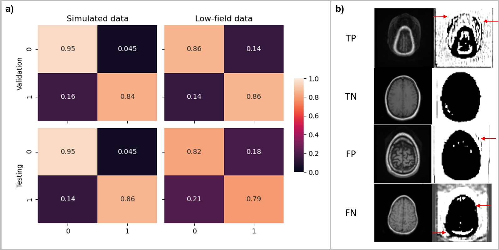
Figure 4. Performance evaluation and model explanation via filter visualization for wrap-around artifact identification. (a) Confusion matrix obtained from the validation and test sets; in the case of simulated data, forward modeling parameters were tweaked based on the validation set results to improve performance. (b) Visualization of representative filters for a true-positive (TP), true-negative (TN), false-positive (FP) and a false-negative (FN) input for explainability.
