Yuning Gu1, Huiyun Gao2, Kihwan Kim1, Yunmei Wang2, and Xin Yu1,3
1Biomedical Engineering, Case Western Reserve University, Cleveland, OH, United States, 2Medicine, Case Western Reserve University, Cleveland, OH, United States, 3Radiology, Case Western Reserve University, Cleveland, OH, United States
1Biomedical Engineering, Case Western Reserve University, Cleveland, OH, United States, 2Medicine, Case Western Reserve University, Cleveland, OH, United States, 3Radiology, Case Western Reserve University, Cleveland, OH, United States
We developed a model-based
approach for CMRO2 mapping in the post-stroke mouse brain with 1.6-mm nominal
resolution at 9.4 T. The method showed
~50% reduced CMRO2 in the infarct core.
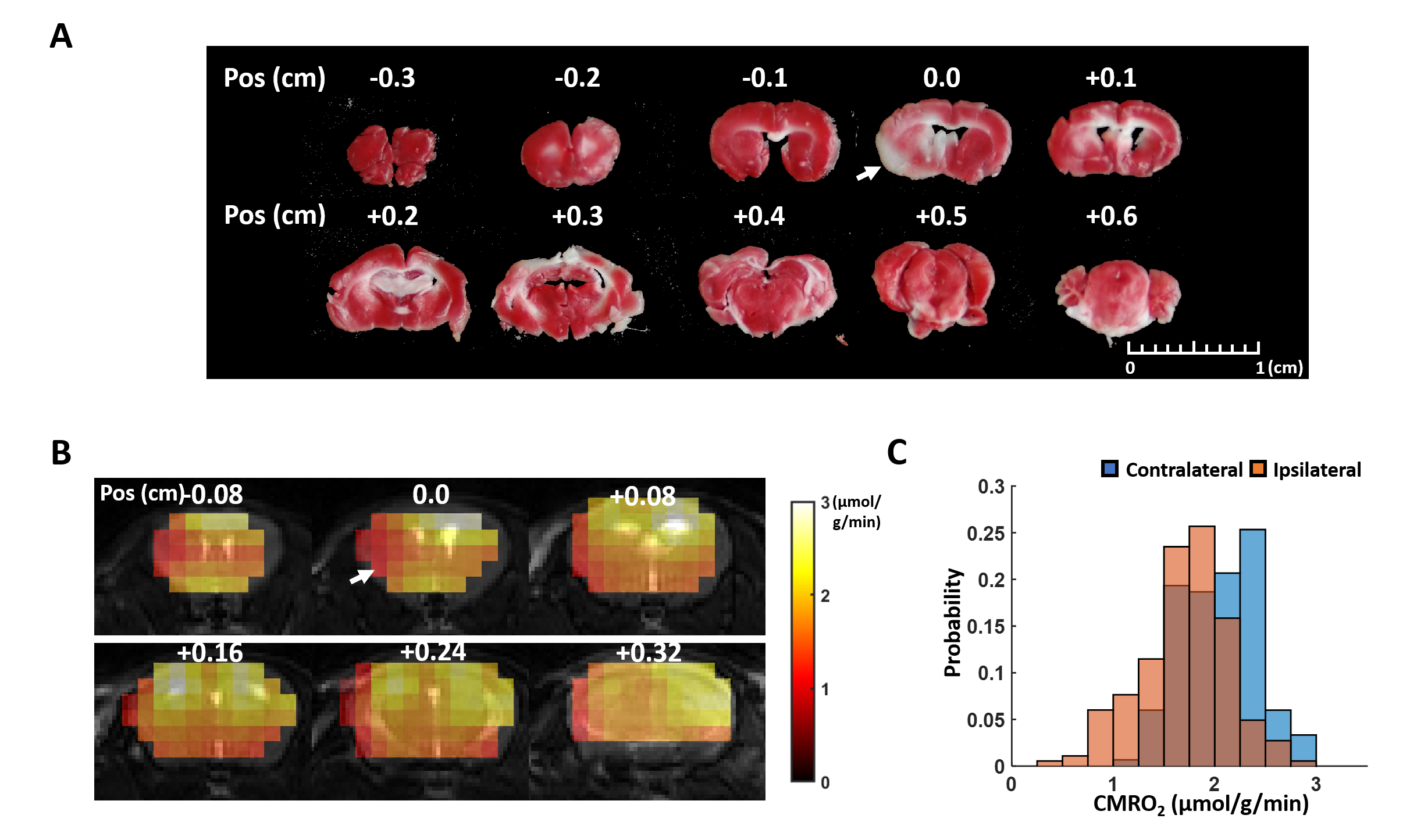
Figure 3. In vivo results. A. TTC staining of the post-stroke mouse
brain. B. CMRO2 maps
overlayed on T2-weighted image.
C. Histogram of CMRO2 values in the ipsilateral and
contralateral hemisphere.
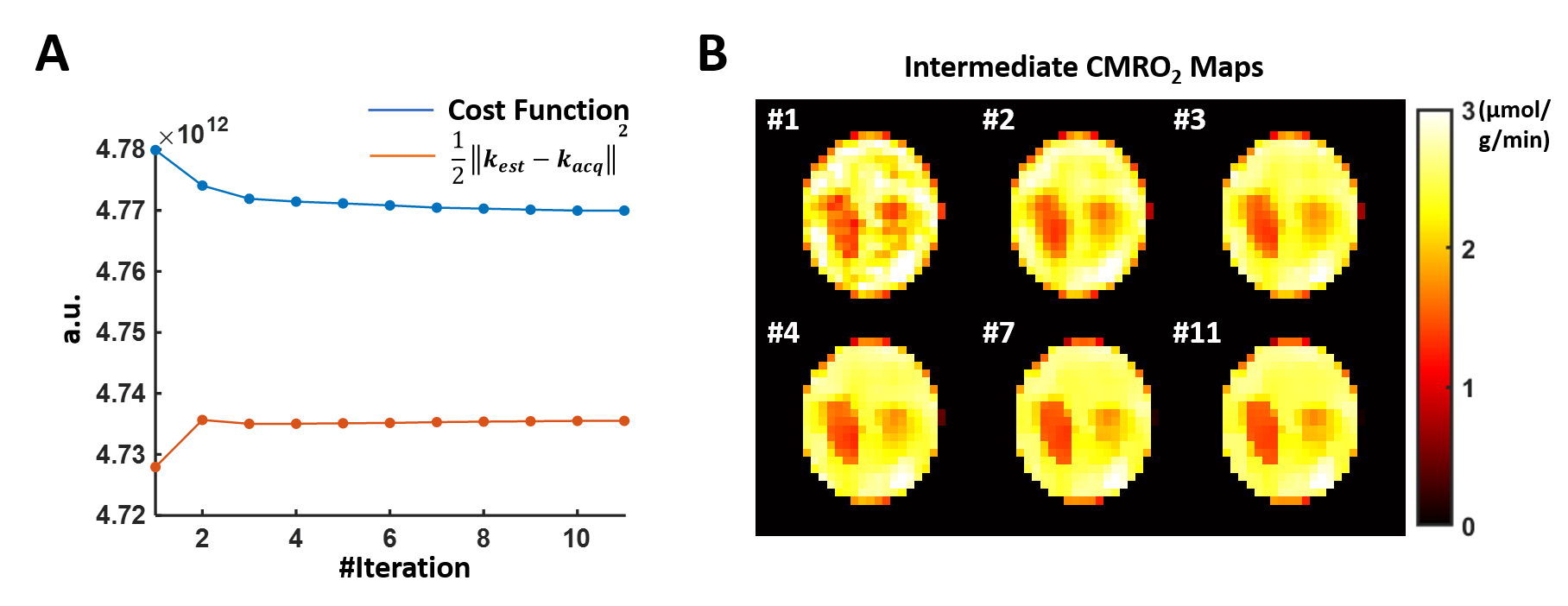
Figure 1. Intermediate results in the
simulation study at high SNR. A. The
entire cost function (blue line) and the error from data consistency term (red
line) in each iteration. B. Intermediate
CMRO2 mapping results in each iteration.