Zhensen Chen1, Yishi Wang2, Le He1, Hanyu Wei1, Yibing Chen3, and Xiaolei Song1
1Center for Biomedical Imaging Research, School of Medicine, Tsinghua University, Beijing, China, 2Philips Healthcare, Beijing, China, 3School of Information Science and Technology, Northwest University, Xi'an, China
1Center for Biomedical Imaging Research, School of Medicine, Tsinghua University, Beijing, China, 2Philips Healthcare, Beijing, China, 3School of Information Science and Technology, Northwest University, Xi'an, China
A
new CEST saturation preparation and readout scheme that allows discrimination
of fast and slow exchange species was proposed, implemented and tested at 3TMR
scanner for human brain imaging.

Fig.3. PaVARS CEST images
and signal evolution for healthy human brain at 3T. A, B and C are
MTRasym images at 3.5ppm, Amide signal and NOE signal using LD quantification,
respectively. Images from the 5 modules
of PaVARS and the first
principal component of the five modules are shown. D: T2w FLARE image showing the drawn ROI of putamen, caudate and globus
pallidus. E shows the five PaVARS Zspectra and the
corresponding Lorentzian fitted lines and the zoomed residual (LD) spectra. F plots the LD signal changes as sat.
module #, with the error bar indicating standard deviation across subjects.
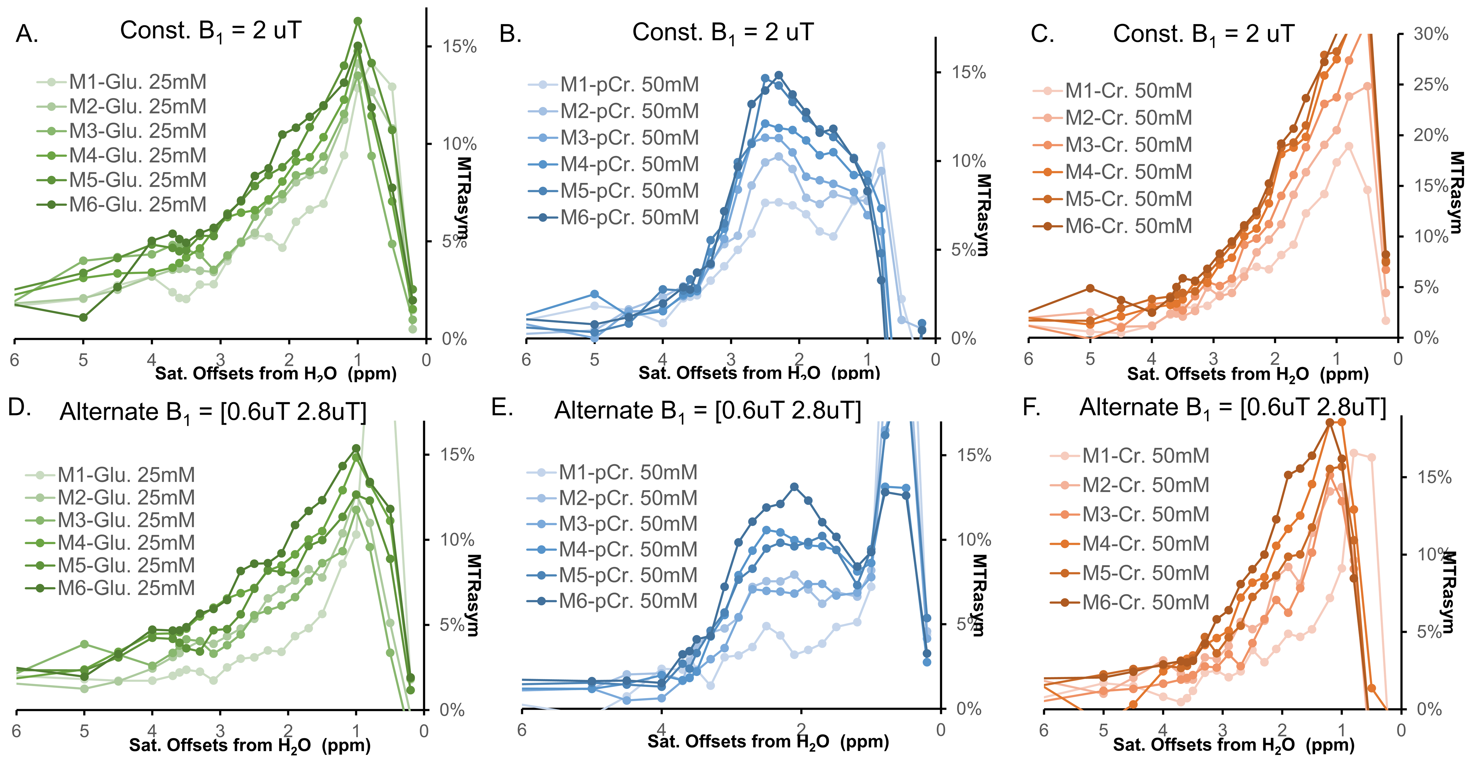
Fig.2. Phantom experiments of PaVARS CEST profile
for three different metabolites, from left to right: Glu., pCr. and Cr. The upper
row was obtained with constant B1 =2uT, while the bottom row was obtained with
an alternating B1 of 0.6uT and 2.8uT. As seen, PaVARS allows a unique profile
for Glu., pCr. and Cr., despite that pCr. and Cr. both contain guanidinium amine
protons and the Glutamate amine resonances at similar offset frequency.