Kay Jann1, Xingfeng Shao1, Samantha J Ma1,2, Karl G Helmer3, Michael Magaletta3, Mitchell J Horn4, Andrew D Warren4, Vanessa A Gonzalez4, Hanzhang Lu5, Yang Li5, Zixuan Lin5, Kaisha Hazel5, George Pottanat5, and Danny JJ Wang1
1Laboratory of Functional MRI Technology (LOFT), USC Stevens Neuroimaging and Informatics Institute, Keck School of Medicine, University of Southern California (USC), Los Angeles, CA, United States, 2Siemens Medical Solutions USA, Inc., Los Angeles, CA, United States, 3Department of Radiology, Massachusetts General Hospital and Athinoula A Martinos Center for Biomedical Imaging, Harvard Medical School, Charlestown, MA, United States, 4Department of Neurology, Massachusetts General Hospital and Athinoula A Martinos Center for Biomedical Imaging, Harvard Medical School, Charlestown, MA, United States, 5The Russell H. Morgan Department of Radiology & Radiological Science, Johns Hopkins University School of Medicine, Baltimore, MD, United States
1Laboratory of Functional MRI Technology (LOFT), USC Stevens Neuroimaging and Informatics Institute, Keck School of Medicine, University of Southern California (USC), Los Angeles, CA, United States, 2Siemens Medical Solutions USA, Inc., Los Angeles, CA, United States, 3Department of Radiology, Massachusetts General Hospital and Athinoula A Martinos Center for Biomedical Imaging, Harvard Medical School, Charlestown, MA, United States, 4Department of Neurology, Massachusetts General Hospital and Athinoula A Martinos Center for Biomedical Imaging, Harvard Medical School, Charlestown, MA, United States, 5The Russell H. Morgan Department of Radiology & Radiological Science, Johns Hopkins University School of Medicine, Baltimore, MD, United States
Standardized 3D background suppressed pCASL scans were performed on a traveling cohort of 10 volunteers. Regional CBF can be reliably estimated across four major MR vendor platforms when accounting for differences in global CBF.
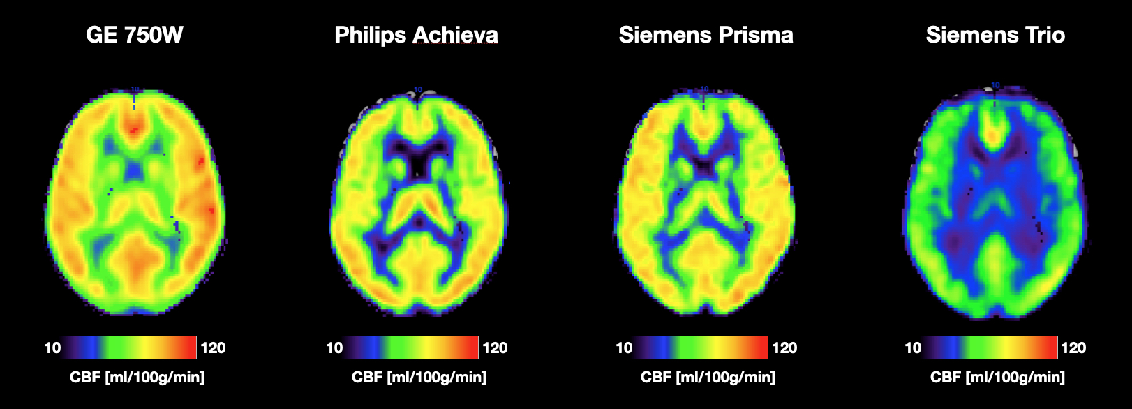
Figure 1: Average CBF maps for all four scanner platforms. All maps are scaled the same which makes the baseline difference in global CBF calculation evident. The perfusion pattern however looks consistent across scanners.
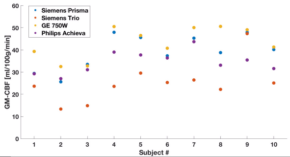
Figure 2: Gray Matter CBF
for each participant and site, highlighting scanner platform bias as
well as covariation of CBF measurements across subjects despite the
global bias.