Lucy V Hiscox1, Emma M Tinney1, Peyton L Delgorio1, Matthew DJ McGarry2, Alyssa Lanzi3, James M Ellison4, Matthew L Cohen3, Chris R Martens5, and Curtis L Johnson1
1Department of Biomedical Engineering, University of Delaware, Newark, DE, United States, 2Dartmouth College, Hanover, NH, United States, 3Department of Communication Sciences & Disorders, University of Delaware, Newark, DE, United States, 4Swank Center for Memory Care and Geriatric Consultation, ChristianaCare, Wilmington, DE, United States, 5Kinesiology and Applied Physiology, University of Delaware, Newark, DE, United States
1Department of Biomedical Engineering, University of Delaware, Newark, DE, United States, 2Dartmouth College, Hanover, NH, United States, 3Department of Communication Sciences & Disorders, University of Delaware, Newark, DE, United States, 4Swank Center for Memory Care and Geriatric Consultation, ChristianaCare, Wilmington, DE, United States, 5Kinesiology and Applied Physiology, University of Delaware, Newark, DE, United States
The health and integrity of the hippocampus is assessed using structural MRI, diffusion imaging, arterial spin labelling, and magnetic resonance elastography (MRE). We find that mechanical property measures, obtained from MRE, are the best predictors of mild cognitive impairment.

Figure 2. Structural MRI and MRE stiffness images of A) a healthy older adult, and B) a patient with mild cognitive impairment (MCI). While smaller volumes of the hippocampus (arrows) are predictive of MCI, viscoelasticity measures from MRE presented a greater risk factor for a clinical diagnosis.
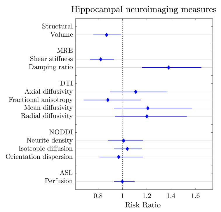
Figure 1. Age and sex adjusted association (risk ratios and 95% CIs) between hippocampal neuroimaging measures and the risk of MCI.