Omar Narvaez1, Maxime Yon2, Alejandra Sierra1, and Daniel Topgaard3
1A.I. Virtanen Institute for Molecular Sciences, University of Eastern Finland, Kuopio, Finland, 2CEMHTI, French National Centre for Scientific Research, Paris, France, 3Department of Chemistry, Lund University, Lund, Sweden
1A.I. Virtanen Institute for Molecular Sciences, University of Eastern Finland, Kuopio, Finland, 2CEMHTI, French National Centre for Scientific Research, Paris, France, 3Department of Chemistry, Lund University, Lund, Sweden
We present
nonparametric D(ω)-distributions as a joint analysis taking
both frequency-dependence and tensorial properties into account, and
demonstrate with ex vivo rat brain data acquired with
gradient waveforms exploring dimensions of the
tensor-valued encoding spectrum b(ω).

Figure 2. Results for representative individual voxels (crosses on
the S(b = 0) map) in an ex vivo rat brain at 90 µm3 isometric
resolution. The D(ω)-distributions, shown as projections onto the 2D
plane of isotropic diffusivity Diso and squared normalized anisotropy
DΔ2 with gray scale of contour lines given by the frequency ω, are
estimated from the b(ω)-encoded signals (circles: measured, points: fit)
by Monte Carlo inversion (45,48). Segmentation into tissue types is
performed by defining bins in the Diso-DΔ2 plane and calculating per-bin
signal fractions fbin1, fbin2, and fbin3.
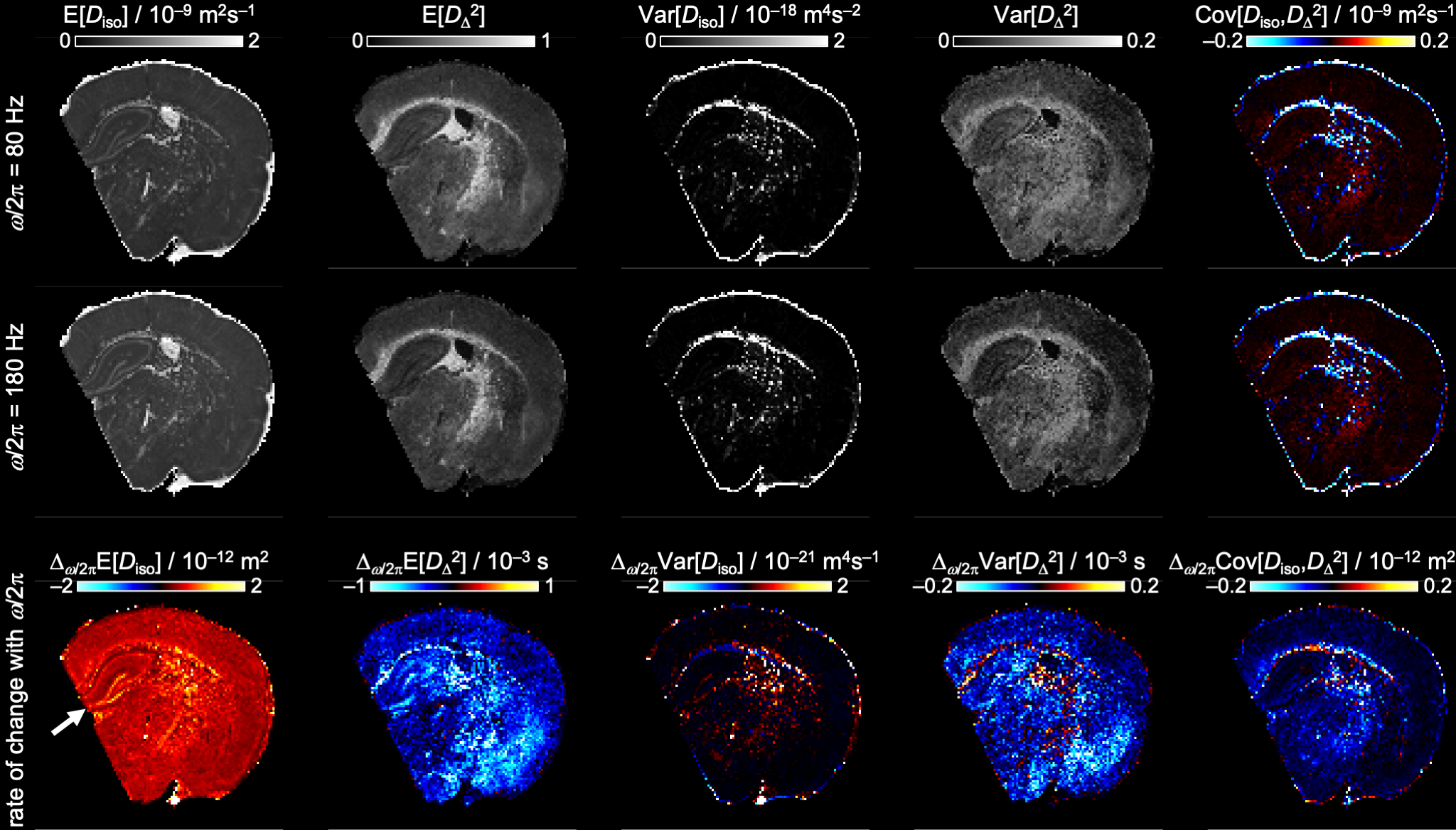
Figure 4. Per-voxel statistical descriptors E[x], Var[x], and
Cov[x,y] over the Diso and DΔ2 dimensions of the D(ω)-distributions for
two selected frequencies ω/2π = 80 and 180 Hz (top and middle rows) and
the rate of change with frequency Δω/2π of the various metrics
(bottom row). The white arrow indicates the hippocampus with elevated
values of Δω/2πE[Diso] in the pyramidal and granule cell layer.