Huaxia Pu1, Haoyao Cao2, Yubo Fan3, Jinge Zhang1, Zhenlin Li1, Zhan Liu2, Liqing Peng1, Tinghui Zheng2, Xiaoyue Zhou4, and Ning Jin5
1Department of Radiology, West China Hospital, Sichuan University, Chengdu, China, 2Department of Applied Mechanics, Sichuan University, Chengdu, China, 3Key Laboratory for Biomechanics and Mechanobiology of the Ministry of Education, School of Biological Science and Medical Engineering, Beihang University, Beijing, China, 4MR Collaboration, Siemens Healthineers Ltd, Shanghai, China, 5Siemens Medical Solutions USA, Chicago, IL, United States
1Department of Radiology, West China Hospital, Sichuan University, Chengdu, China, 2Department of Applied Mechanics, Sichuan University, Chengdu, China, 3Key Laboratory for Biomechanics and Mechanobiology of the Ministry of Education, School of Biological Science and Medical Engineering, Beihang University, Beijing, China, 4MR Collaboration, Siemens Healthineers Ltd, Shanghai, China, 5Siemens Medical Solutions USA, Chicago, IL, United States
Helical flow exists in normal diastolic superior vena cava flow. Flow pathlines from brachiocephalic veins form these helixes,
resulting in two types: twining and untwining.
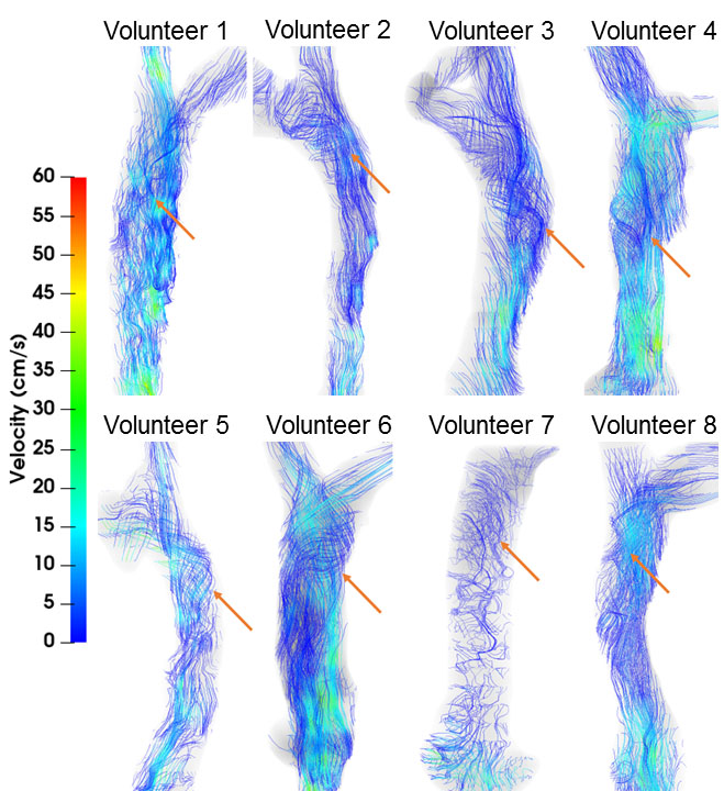
Figure 1. Streamline visualizations (color-coded to blood speed) in all 8
subjects. Helical flow (red arrows) was observed in all subjects.

Figure 3. Superior vena cava pathline visualizations in 4
volunteers at three cardiac timepoints each, showing (from
left to right) no helical flow, matured helical flow, and disrupted helical
flow, respectively. Type 1 (twining) is shown in the top row, and Type 2
(untwining) in the bottom row.