Qian Wan1,2, Hao Peng 1, Jianxun Lyu1,2, Xiaoyi Liu3, Chuanli Cheng1, Feng Liu3, Yangzi Qiao1, Hairong Zheng1, Xin Liu1, Yi Wang3, and Chao Zou1
1Shenzhen Institutes of Advanced Technology, Chinese Academy of Sciences, Shenzhen, China, 2Shenzhen College of Advanced Technology,University of Chinese Academy of Sciences, Shenzhen, China, 3Peking University People's Hospital, Beijing, China
1Shenzhen Institutes of Advanced Technology, Chinese Academy of Sciences, Shenzhen, China, 2Shenzhen College of Advanced Technology,University of Chinese Academy of Sciences, Shenzhen, China, 3Peking University People's Hospital, Beijing, China
We detected the liver fibrosis using water specific T1 mapping with Gadoxetic Acid -enhanced MRI in a rat NASH model and revealed that the reduction rates of water-T1 relaxation time after Gd-EOB-DTPA administration might be a useful tool to stage the mild and moderate live fibrosis in rat model.
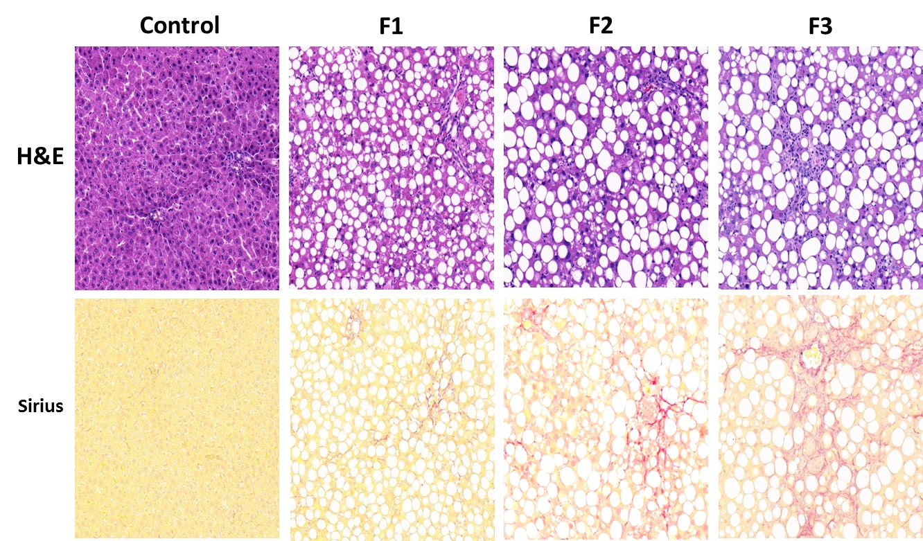
Fig. 1. Typical histological sections of the
control and
MCD diet groups
by Hematoxylin-eosin
(HE)
and Sirius red staining.
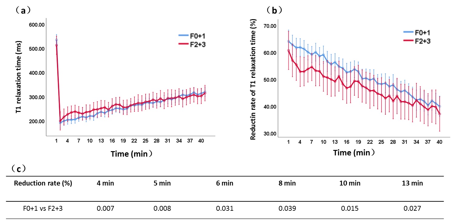
Fig. 2. Line graphs of T1 relaxation time (a) and
reduction rate of T1 relaxation time of the liver (b)
in different stage of fibrosis.
(c)
Adjusted P
Value of Reductin rate of T1 relaxation time (%) F0+1 vs
F2+3.