Kadi Vaher1, Paola Galdi1, Manuel Blesa Cabez1, Gemma Sullivan1, Gill Black1, David Q Stoye1, Alan J Quigley2, Michael J Thrippelton3, Simon R Cox4, Mark E Bastin3, Debby Bogaert5, and James P Boardman1
1MRC Centre for Reproductive Health, University of Edinburgh, Edinburgh, United Kingdom, 2Department of Paediatric Radiology, Royal Hospital for Sick Children, Edinburgh, United Kingdom, 3Centre for Clinical Brain Sciences, University of Edinburgh, Edinburgh, United Kingdom, 4Lothian Birth Cohort Studies group, Department of Psychology, University of Edinburgh, Edinburgh, United Kingdom, 5Centre for Inflammation Research, University of Edinburgh, Edinburgh, United Kingdom
1MRC Centre for Reproductive Health, University of Edinburgh, Edinburgh, United Kingdom, 2Department of Paediatric Radiology, Royal Hospital for Sick Children, Edinburgh, United Kingdom, 3Centre for Clinical Brain Sciences, University of Edinburgh, Edinburgh, United Kingdom, 4Lothian Birth Cohort Studies group, Department of Psychology, University of Edinburgh, Edinburgh, United Kingdom, 5Centre for Inflammation Research, University of Edinburgh, Edinburgh, United Kingdom
Applying PCA to tract-averaged diffusion MRI metrics reveals
substantial shared variance within and between DTI and NODDI metrics across 16
major white matter tracts in a neonatal population. This property enables
derivation of general factors, which associate with preterm birth.
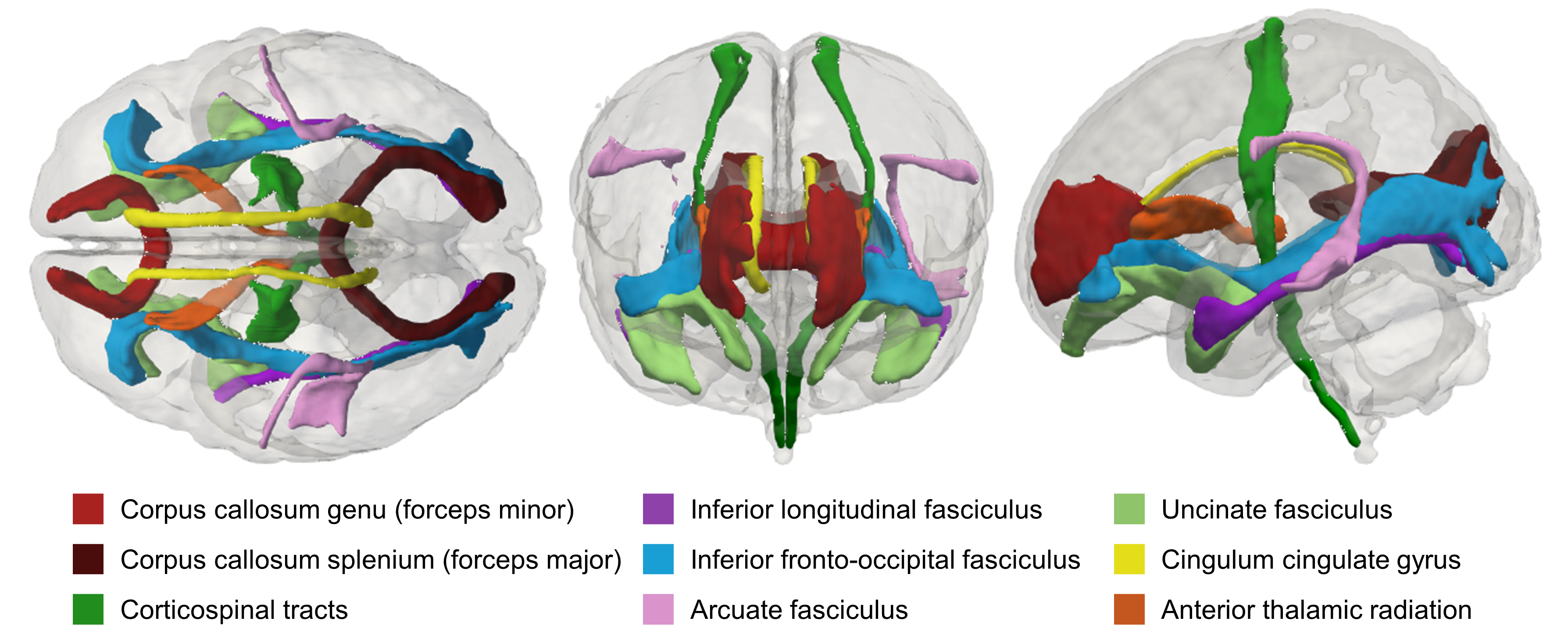
Figure 1. Visual
representation of the delineated white matter tracts in the neonatal atlas
space. Shown in superior (left), anterior (centre) and lateral (right) views.
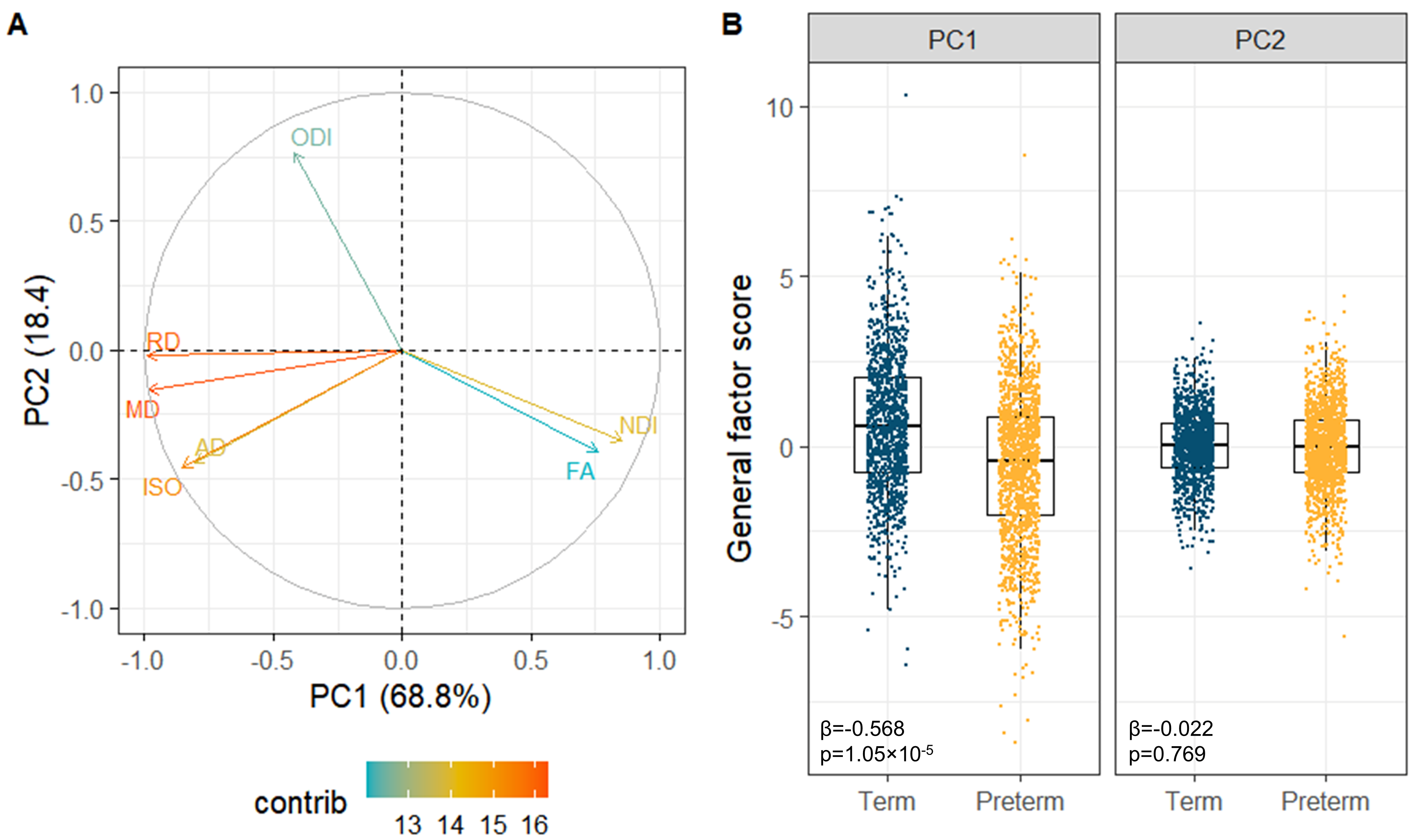
Figure 3. Multimodal
general factors. (A) PCA variable contribution plot; the colours represent the
contribution of the dMRI metric to the components. (B) Boxplots of the
multimodal g-factors showing differences between the term and preterm group.
Note that each participant is represented by 16 tracts. Reported statistics are
standardised β for the PC1 or
PC2 from linear mixed effect regression models and FDR-corrected p-values.