Jérémie Clément1, Kathleen Colford2, Emer Hughes2, Tomoki Arichi2,3,4, David Edwards2,3,5, Joseph Hajnal1,2, and Ozlem Ipek1
1Biomedical Engineering, Kings College London, London, United Kingdom, 2Centre for the developing brain, Kings College London, London, United Kingdom, 3Bioengineering, Imperial College London, London, United Kingdom, 4Paediatric neurosciences, Evelina London children's hospital, Guy's and St Thomas' NHS Foundation Trust, London, United Kingdom, 5MRC Centre for Neurodevelopmental Disorders, Kings College London, London, United Kingdom
1Biomedical Engineering, Kings College London, London, United Kingdom, 2Centre for the developing brain, Kings College London, London, United Kingdom, 3Bioengineering, Imperial College London, London, United Kingdom, 4Paediatric neurosciences, Evelina London children's hospital, Guy's and St Thomas' NHS Foundation Trust, London, United Kingdom, 5MRC Centre for Neurodevelopmental Disorders, Kings College London, London, United Kingdom
In this work, we
describe the first mechanical frame for infants for brain and cardiac imaging
at 7T. The different parts incorporate important design aspects including mechanical
sturdiness/safety, toxicity, sanitization and ease of use.
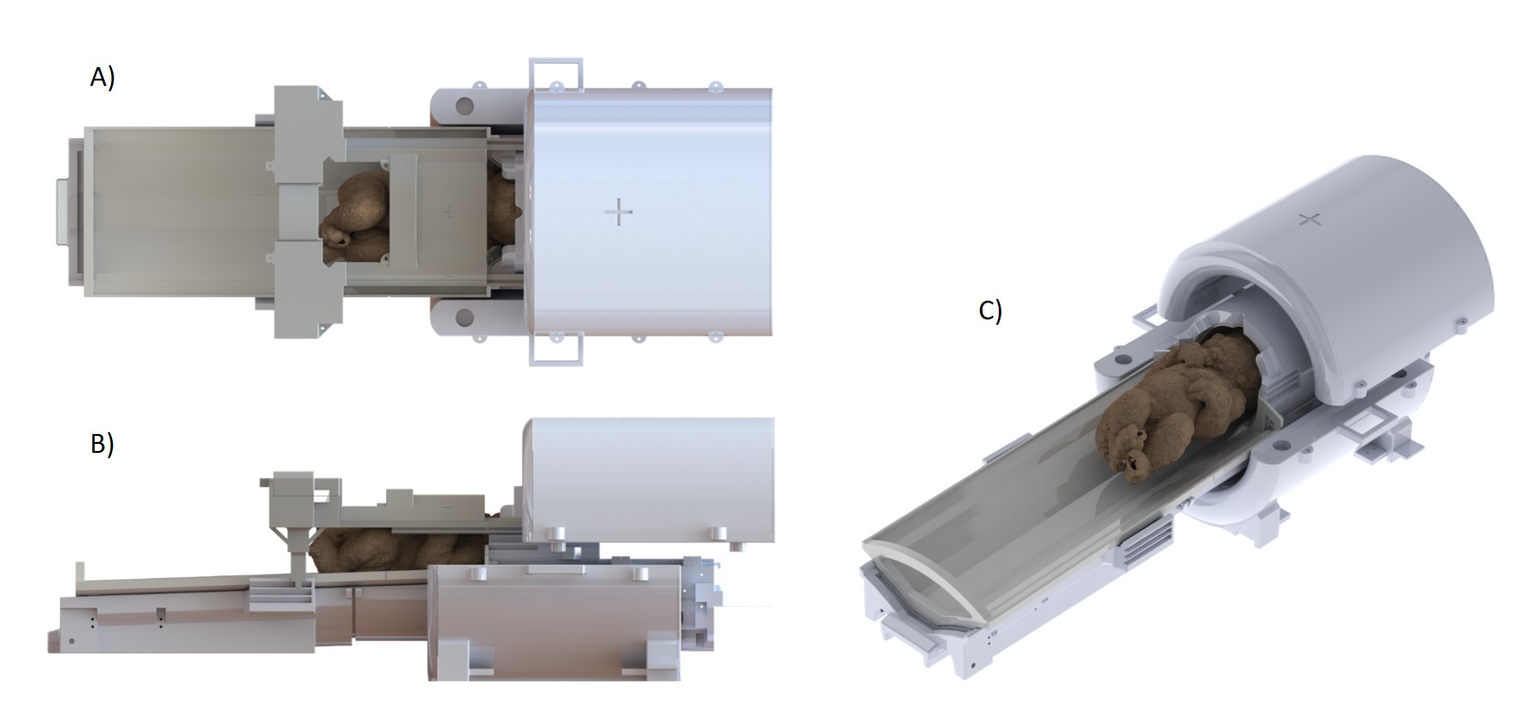
Figure
4 – A) and B) 3D render of the structural elements from Fig. 1A-D. A infant
model11 was added for better visualization of the positioning. The Tx-array
coil holder was slightly moved out of the final position. C) 3D render of the
structural elements from Fig. 1A-C. The fitting of the baby model can be
clearly evaluated. The main support structure (Fig. 1E) is not shown to
simplify the view.
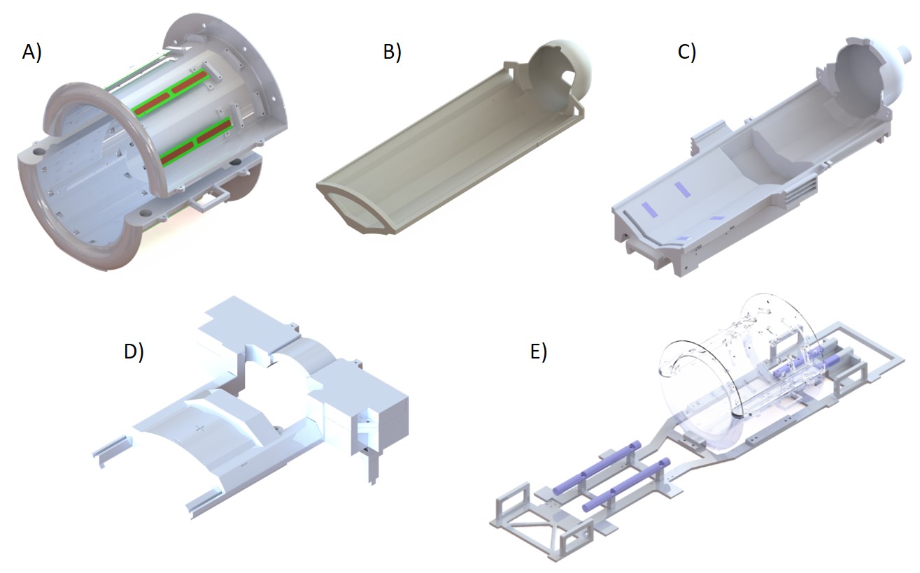
Figure 1 - 3D render of the main parts of the mechanical frame for infants at 7T, with
A) the Tx-array
coil holder showing the position of the dipole elements, B) the bed structure,
C) the posterior Rx-array frame, D) the anterior Rx-array frame and E) the main
support structure. The parts displayed in light blue (in C and E) correspond to
the Teflon pads used for displacement between the brain-isocentred and
heart-isocentred positions.