Yuhui Liu1, AIlian Liu1, Anliang Chen1, Jiazheng Wang2, Geli Hu2, Wan Dong1, Qingwei Song1, Mingxiao Wang1, Xinru Zhang1, and Xinao Wang1
1The First Affiliated Hospital of Dalian Medical University, Dalian, China, Dalian, China, 2Philips Healthcare,Beijing,China, Beijing, China
1The First Affiliated Hospital of Dalian Medical University, Dalian, China, Dalian, China, 2Philips Healthcare,Beijing,China, Beijing, China
T2 mapping can be used as a non-invasive technique to distinguish rectal tubular adenocarcinoma from non-tubular adenocarcinoma.
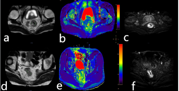
Figure 1 A 77-year-old
male patient with the rectal tubular adenocarcinoma. T2 image (a), T2 mapping
image (b), and DWI image (c) were showed. A 63-year-old male patient with rectal
non-tubular adenocarcinoma. X image (d), T2 mapping image (e), and X image (f)
were showed.
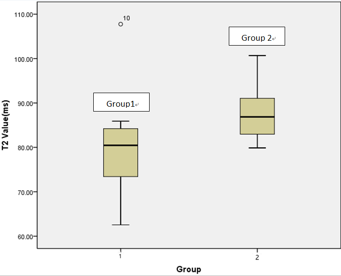
Figure2 T2 values
of rectal tubular adenocarcinoma (group1) and non-tubular adenocarcinoma
(group2) with a significant difference observed (p =0.041).