Yajie Wang1, Ming Xiao2, Canhong Xiang2, Yuewei Zhang2, Haikun Qi3, Yishi Wang4, Jiahong Dong2, and Huijun Chen1
1Center for Biomedical Imaging Research, Department of Biomedical Engineering, School of Medicine, Tsinghua University, Beijing, China, 2Hepato-pancreato-biliary Center, Beijing Tsinghua Changgung Hospital, School of Medicine, Tsinghua University, Beijing, China, 3School of Biomedical Engineering and Imaging Sciences, King's College London, London, United Kingdom, 4Philips Healthcare, Beijing, China
1Center for Biomedical Imaging Research, Department of Biomedical Engineering, School of Medicine, Tsinghua University, Beijing, China, 2Hepato-pancreato-biliary Center, Beijing Tsinghua Changgung Hospital, School of Medicine, Tsinghua University, Beijing, China, 3School of Biomedical Engineering and Imaging Sciences, King's College London, London, United Kingdom, 4Philips Healthcare, Beijing, China
A
free-breathing whole liver T1 mapping technique was achieved in a single scan
using GOAL-SNAP sequence. The potential of the proposed technique for liver
function estimation was demonstrated in patients with hilar cholangiocarcinoma.
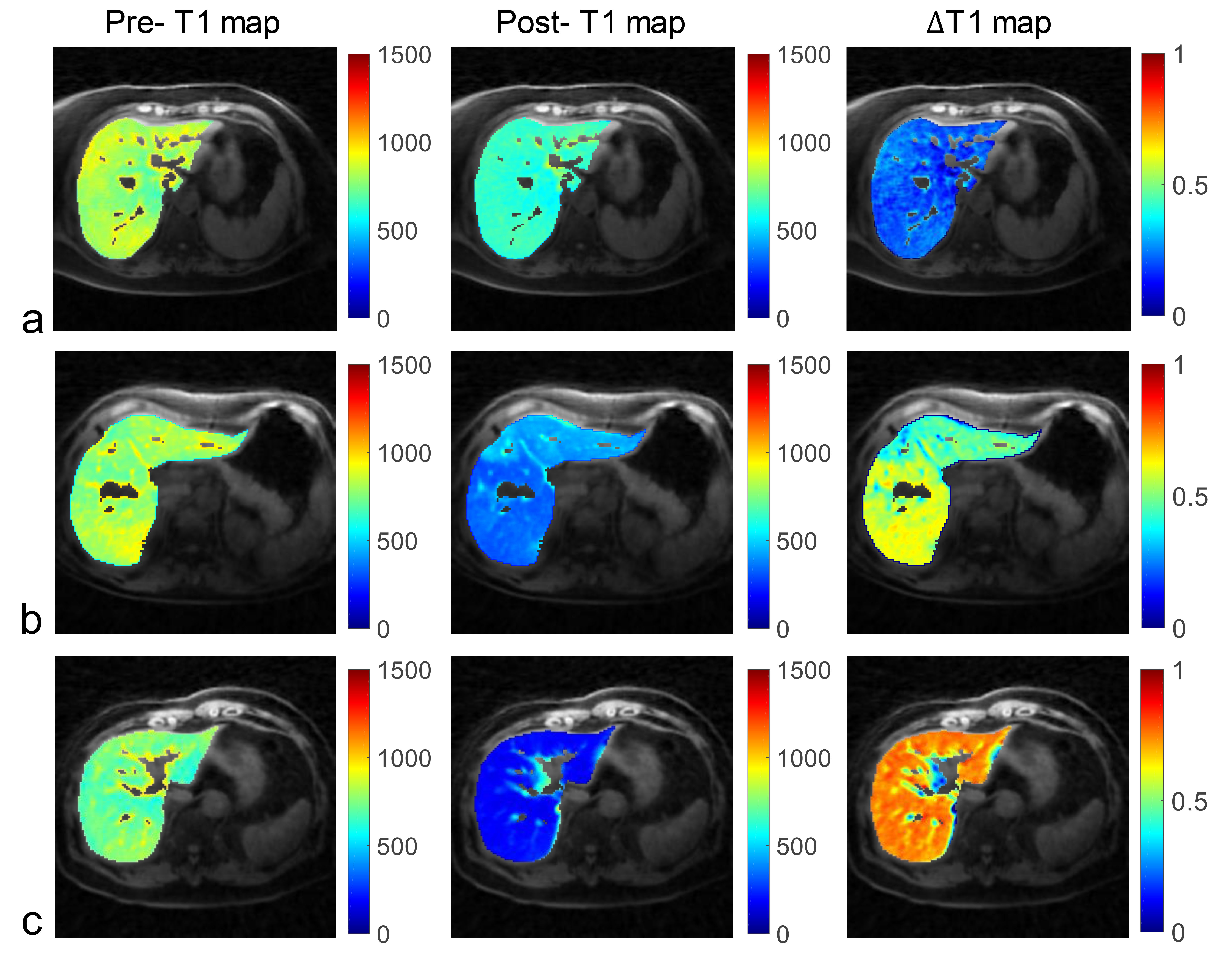
Figure
3. Pre-contrast T1 map, post-contrast T1 map and ∆T1
map of three patients with hilar
cholangiocarcinoma
(HCCA). (a) Maps of one patient (female, age
62 years) acquired after bilateral drainage;
(b)
Maps of one patient (female, age 64 years) acquired after bilateral drainage
and left portal vein
embolization (PVE); (c)
Maps of one patient (female, age
67 years) without drainage
and PVE.
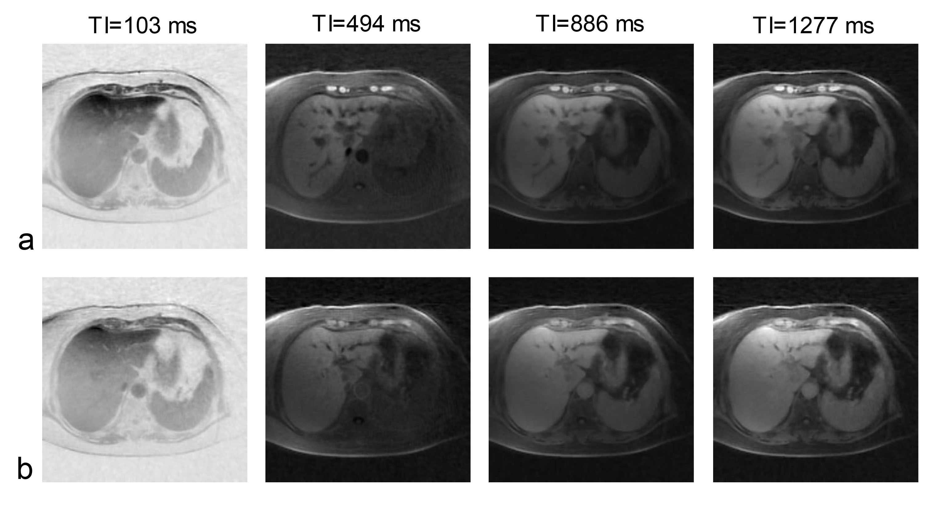
Figure
2. Pre-contrast (a) and 20 min post-contrast (b) T1-weighted images at
different inversion times (TI) reconstructed from GOAL-SNAP sequence of one
patient (female, 62 years)
with hilar cholangiocarcinoma
(HCCA).