Lihua Chen1, Ailian Liu1, Qingwei Song1, and Lizhi Xie2
1The First Affiliated Hospital of DaLian Medical University, DaLian , China., Dalian, China, 2GE Healthcare China, Beijing, China, Beijing, China
1The First Affiliated Hospital of DaLian Medical University, DaLian , China., Dalian, China, 2GE Healthcare China, Beijing, China, Beijing, China
So,
it is very important to accurately distinguish between these two tumors before
therapy is planned. The purpose of this study is to evaluate and compare the FA
value of DTI and the ADC value of DWI in ICC and HCC.
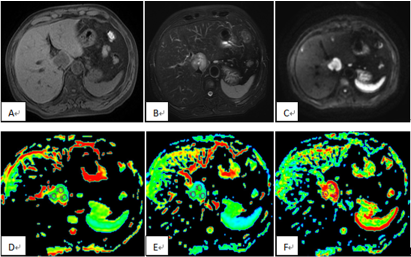
Fig
1: Patient, male,
57 years old, ICC in the caudate lobe of liver. A-C: shows the T1W image (hypointense), T2W image
(hyperintense), and DWI image (hyperintense) respectively. D-F: shows ADC image
of DWI, D, and FA image of DTI respectively. The ADC, D, and FA values were 1.155×10⁻⁹m2/s,
1.465×10⁻⁹m2/s, and 0.571.

Fig
2: Patient, male,
62 years old, HCC in the right lobe of liver. A-C: shows the T1W image
(hypointense), T2W image (hyperintense), and DWI image (hyperintense)
respectively. D-F: shows ADC image of DWI, D, and FA image of DTI respectively.
The ADC, D and FA values were 1.350×10⁻⁹m2/s, 1.655×10⁻⁹m2/s, and 0.399.