Andor Veltien1, Sjaak van Asten1, Nia Ravichandran1, Robin de Graaf2, Henk de Feyter2, Jeannette Oosterwijk3, Egbert Oosterwijk3, and Arend Heerschap1
1Medical Imaging, Radboud UMC, Nijmegen, Netherlands, 2Radiology and biomedical imaging, Yale University, New Haven, CT, United States, 3Urology, Radboud UMC, Nijmegen, Netherlands
1Medical Imaging, Radboud UMC, Nijmegen, Netherlands, 2Radiology and biomedical imaging, Yale University, New Haven, CT, United States, 3Urology, Radboud UMC, Nijmegen, Netherlands
It is possible to follow
the uptake and to image the presence of [2H9]choline in
tumors after a bolus administration of this compound. DMI can be performed simultaneously
of [2H9]choline and of [6,6 2H2]glucose.

Figure 1.
a) 2H MR spectra
of subcutaneous tumor after injection of 2H9 choline.
b)Time curves of HOD and 2H9
choline signal integrals
c) T2
weighted MRI of subcutaneous tumor. d) 2H9 choline heat
map overlaid on T2 weighted MRI
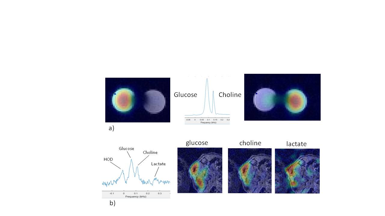
Figure 2
a) DMI of phantom with two tubes, one filled with 2H9 choline and the other with
[6,6 2H2]glucose. The glucose and choline signals are
clearly separated in the 2H spectrum
b) Left: 2H spectrum of renal tumor
after IV bolus infusion of 2H9 choline and [6,6 2H2]glucose
combined in the tail vein of a mouse. Right DMI maps of deuterated glucose,
choline and lactate overlaid on T2 MRI