Yoshitaka Bito1,2, Hisaaki Ochi1,2, Kuniaki Harada2, and Kohsuke Kudo2
1Healthcare Business Unit, Hitachi, Ltd., Tokyo, Japan, 2Department of Diagnostic Imaging, Hokkaido University Graduate School of Medicine, Sapporo, Japan
1Healthcare Business Unit, Hitachi, Ltd., Tokyo, Japan, 2Department of Diagnostic Imaging, Hokkaido University Graduate School of Medicine, Sapporo, Japan
Low b-value DTI was measured to investigate
pseudo-random flow of CSF for normal volunteers. The measured diffusion properties
show significantly high and anisotropic values in some regions; for instance,
around the aqueduct.

Figure 2. Representative
DTLs at the entrance of fourth ventricle from the aqueduct of two
volunteers: ROI-overlaid images (a) and ellipsoid-representation maps (b).
Yellow boxes and green segments on the images (a) display the regions for
ellipsoid-representation and for statistical analysis, respectively. DTL
is represented as an ellipsoid of which maximum ADC is 20×10-9 m2/s
for each voxel in the maps (b).
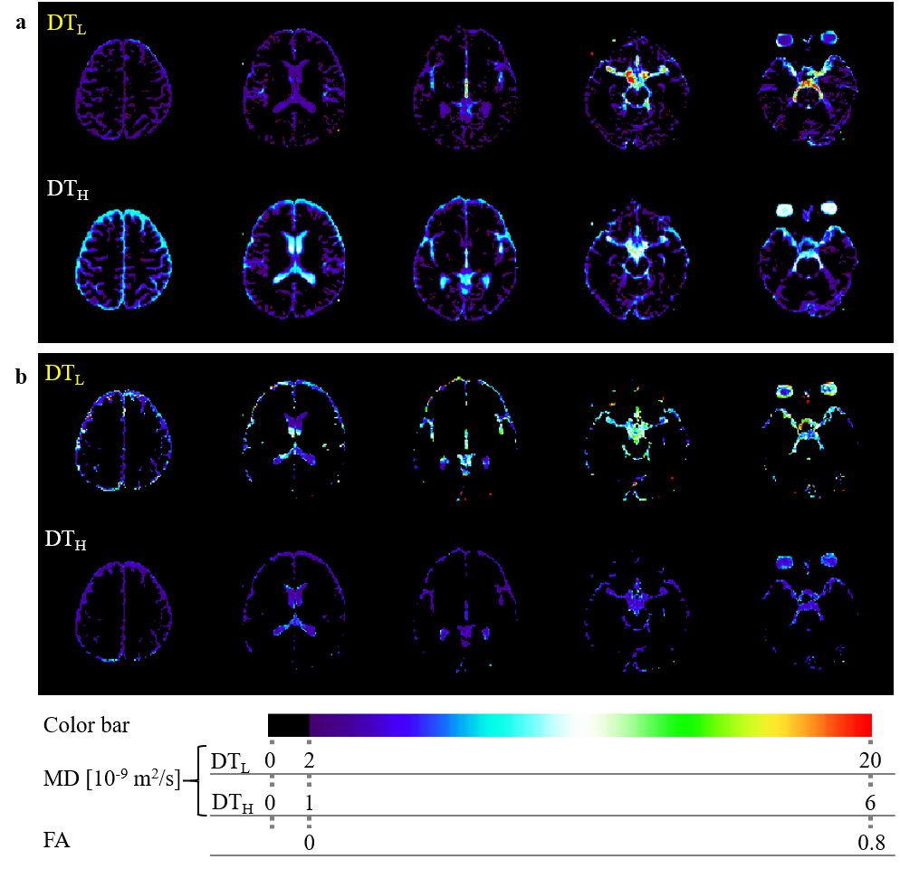
Figure 1. Representative
multislice images of MD (a) and FA (b) of DTL and DTH. DTL
shows extremely high MD and FA in some subsegments of CSF.