Qiyuan Tian1,2, Ziyu Li3, Qiuyun Fan1,2, Chanon Ngamsombat1, Yuxin Hu4, Congyu Liao1,2, Fuyixue Wang1,2, Kawin Setsompop1,2, Jonathan R Polimeni1,2, Berkin Bilgic1,2, and Susie Y Huang1,2
1Athinoula A. Martinos Center for Biomedical Imaging, Massachusetts General Hospital, Boston, MA, United States, 2Harvard Medical School, Boston, MA, United States, 3Department of Biomedical Engineering, Tsinghua University, Beijing, China, 4Department of Electrical Engineering, Stanford University, Stanford, CA, United States
1Athinoula A. Martinos Center for Biomedical Imaging, Massachusetts General Hospital, Boston, MA, United States, 2Harvard Medical School, Boston, MA, United States, 3Department of Biomedical Engineering, Tsinghua University, Beijing, China, 4Department of Electrical Engineering, Stanford University, Stanford, CA, United States
- 1. Proposed a deep learning-based super-resolution method for DTI.
- 2. Employed a very deep convolutional neural network, residual learning, and multi-contrast imaging.
- 3. High-quality results similar to high-resolution ground truth and superior to those from image interpolation.
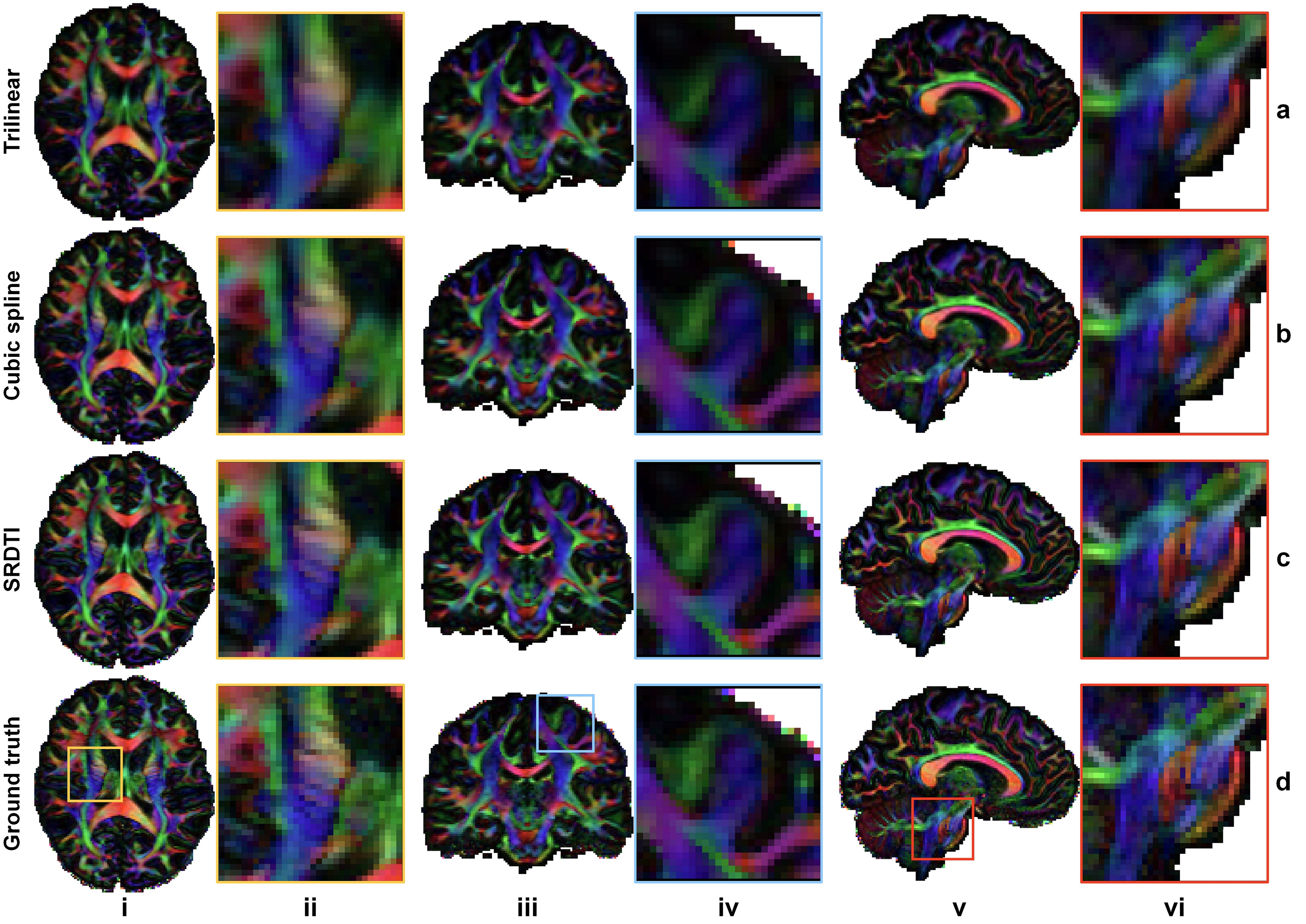
Figure 3. Direction-encoded
fractional anisotropy maps. Fractional anisotropy maps color
encoded by the primary eigenvector (red: left–right; green: anterior–posterior;
blue: superior–inferior) derived from the diffusion tensors fitted using interpolated
data (a, b), SRDTI super-resolution data (c), and ground-truth high-resolution
data (d) along axial, coronal and sagittal directions, showing
regions-of-interest in the internal capsule (i, ii), cerebral cortex (iii, iv)
and pons (v, vi), respectively.
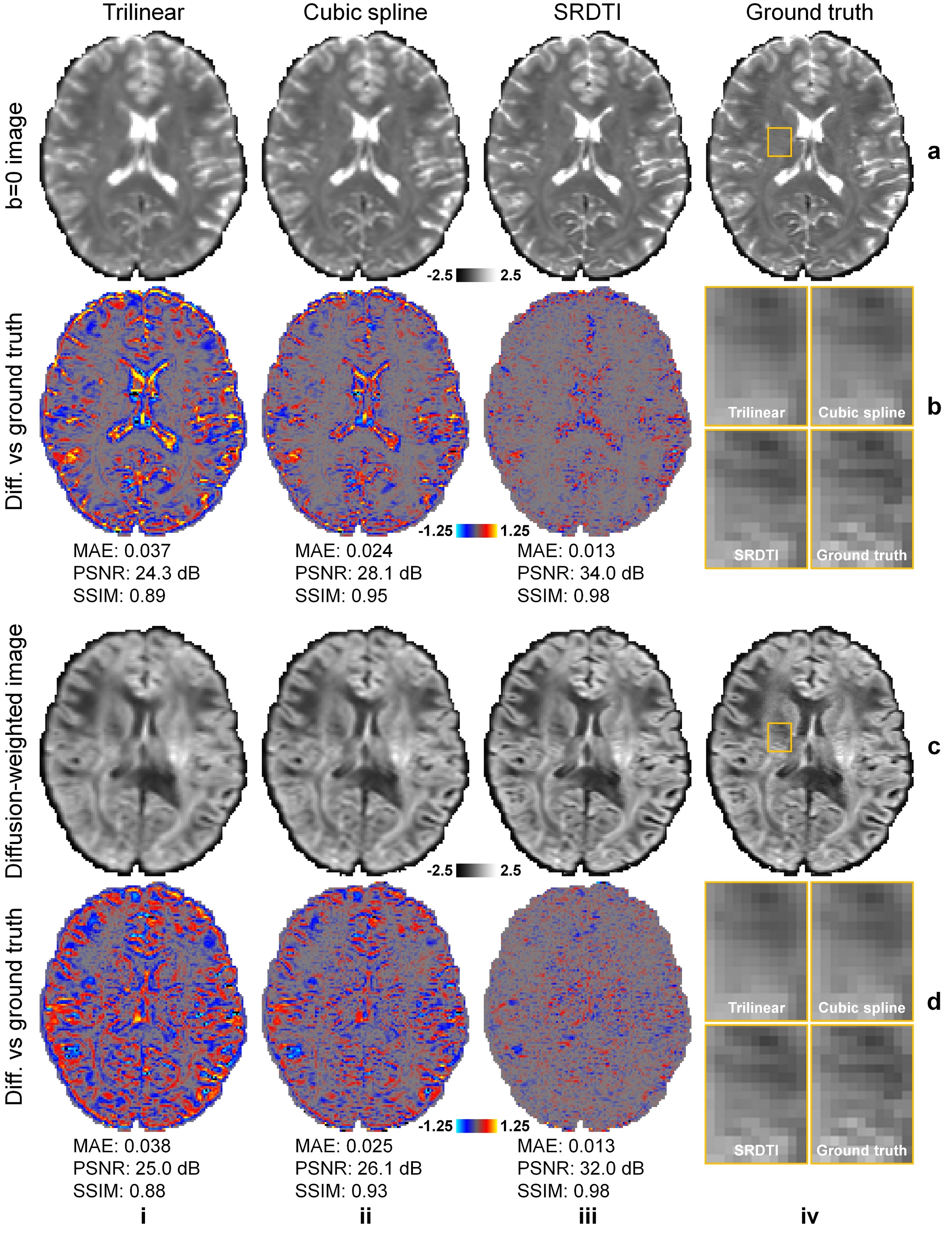
Figure 2. Image results. b=0 images (row a) and diffusion-weighted images (DWIs) (row
c) along one direction of the six optimized diffusion-encoding directions (i.e.,
[0.91, 0.416, 0]) from interpolated data (i, ii) (interpolated data using cubic
spline is the input to SRDTI), SRDTI output data (iii), ground-truth
high-resolution data (iv), and their residuals comparing to the ground-truth high-resolution
images (rows b, d). A region-of-interest in the deep white matter (yellow
boxes) is displayed in enlarged views (rows, b, d, column iv).