Rafael Neto Henriques1, Sune Nørhøj Jespersen2,3, and Noam Shemesh1
1Champalimaud Research, Champalimaud Centre for the Unknown, Lisbon, Portugal, 2Center of Functionally Integrative Neuroscience (CFIN) and MINDLab, Clinical Institute, Aarhus University, Aarhus, Denmark, 3Department of Physics and Astronomy, Aarhus University, Aarhus, Denmark
1Champalimaud Research, Champalimaud Centre for the Unknown, Lisbon, Portugal, 2Center of Functionally Integrative Neuroscience (CFIN) and MINDLab, Clinical Institute, Aarhus University, Aarhus, Denmark, 3Department of Physics and Astronomy, Aarhus University, Aarhus, Denmark
Correation Tensor Imaging revealed that positive sources of intra-compartmental kurtosis bias tensor-valued MDE estimates even in the absence of detectable diffusion
time dependence. A regime in which tensor-valued methods can accurately estimate anisotropic kurtosis is identified.
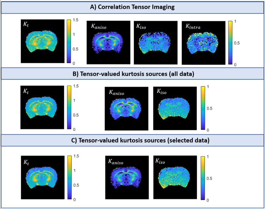
Fig.4 – Kurtosis estimates of a representative rat
brain coronal slice: A) CTI
kurtosis estimates; B) Tensor-valued kurtosis estimates obtained by fitting
Eq. 2 to all acquired data (TV(all)); C) Tensor-valued kurtosis estimates
obtained by fitting Eq. 2 to only the data acquired with b1=b2 and parallel/perpendicular gradient directions (TV(sel)).
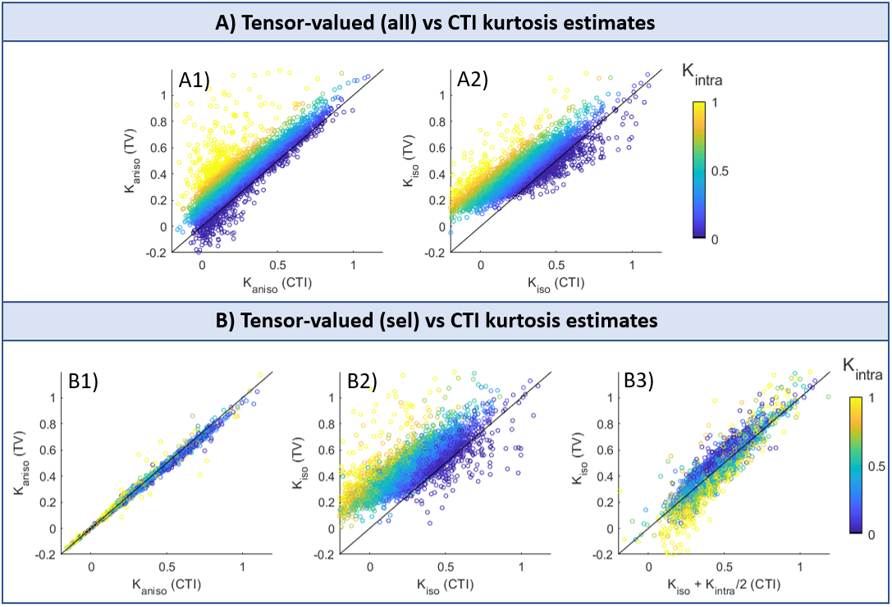
Fig.5 – Scatter plots of tensor-valued vs CTI kurtosis
estimates: A) Tensor-valued kurtosis estimates
obtained by fitting Eq. 2 to all acquired data (TV(all)) vs CTI estimates; B) Tensor-valued
kurtosis estimates obtained by fitting Eq. 2 to DDE data acquired with b1=b2
(TV(sel)) vs CTI estimates. The points in the scatter plots are color-coded according to CTI’s Kintra
estimates.