Yan Ren1, Lihua Chen1, Yizhong Yuan2, Jipan Xu2, Jinxia Zhu3, Robert Grimm4, and Wen Shen1
1Tianjin First Center Hospital, Tianjin, China, 2First Central Hospital Institute, Tianjin Medical University, Tianjin, China, 3MR collaborations, Siemens Healthcare Ltd., Beijing, China, 4Siemens Healthcare GmbH, Erlangen, Germany
1Tianjin First Center Hospital, Tianjin, China, 2First Central Hospital Institute, Tianjin Medical University, Tianjin, China, 3MR collaborations, Siemens Healthcare Ltd., Beijing, China, 4Siemens Healthcare GmbH, Erlangen, Germany
Kidney microcirculation perfusion was the major factor affecting
CIRI, and IVIM imaging may be useful for detecting the renal injury, compensation,
and recoverability.
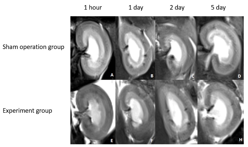
Figure 1. T2WI images acquired at 1
hour (A, E), and at 1 (B, F), 2 (C, G), and 5 (D, H) days after surgery. Figures
A-D are sham operation group. Figures E-H are experiment group.
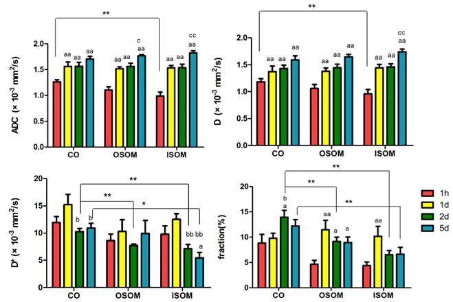
Figure 4. Experimental group graphs
at all imaging time points following the cold ischemia-reperfusion injury
model. Values of the cortex (CO), outer stripe of outer medulla (OSOM), and
inner stripe of outer medulla (ISOM) are averaged over the kidney to give (A)
apparent diffusion coefficients [ADC], (B) pure molecular diffusion [D], (C)
pseudo-diffusion [D*], and (D) the perfusion fraction.
a: versus 1h; b: versus 1d; c:
versus 2d; single mark: P<0.05; double mark: P<0.01;
*: P<0.05; **: P<0.01