Li Liu1, Stephen Dodd1, Ryan Hunt1, Nikorn Pothayee1, Nadia Nadia Bouraoud1, Dragan Maric1, E Ashley Moseman1, Dorian B McGavern1, and Alan P Koretsky1
1National Institute of Neurological Disorders and Stroke, National Institute of Health, Bethesda, MD, United States
1National Institute of Neurological Disorders and Stroke, National Institute of Health, Bethesda, MD, United States
Using microbleeds as an MRI marker of neuroinflammation the time course
of Vesicular Stomatitis Virus nasal infection of the mouse brain was monitored.
Adoptive transfer of virus
specific CD8 T cells reduced brain bleeding. A method to track T cells by MRI
was developed.

Figure
2. Adoptive transfer of CD8 T cells decreased brain bleeding and cleared virus.
MR images of the turbinates (A), OB (B), and brain (C) from non-treated
and CD8 T cell treated mice on 6 (upper
panel) and 11-dpi (lower
panel). (D-F) Quantification of hypointensity spots revealed that CD8 T
cells transfer protected brain vessel integrity. (G) CD8 T cells reduced viral
titers on 6-dpi. (H-I) IHC study revealed that CD8 T cells infiltrated and
proliferated in the GL (H) and core (I) of the OB.
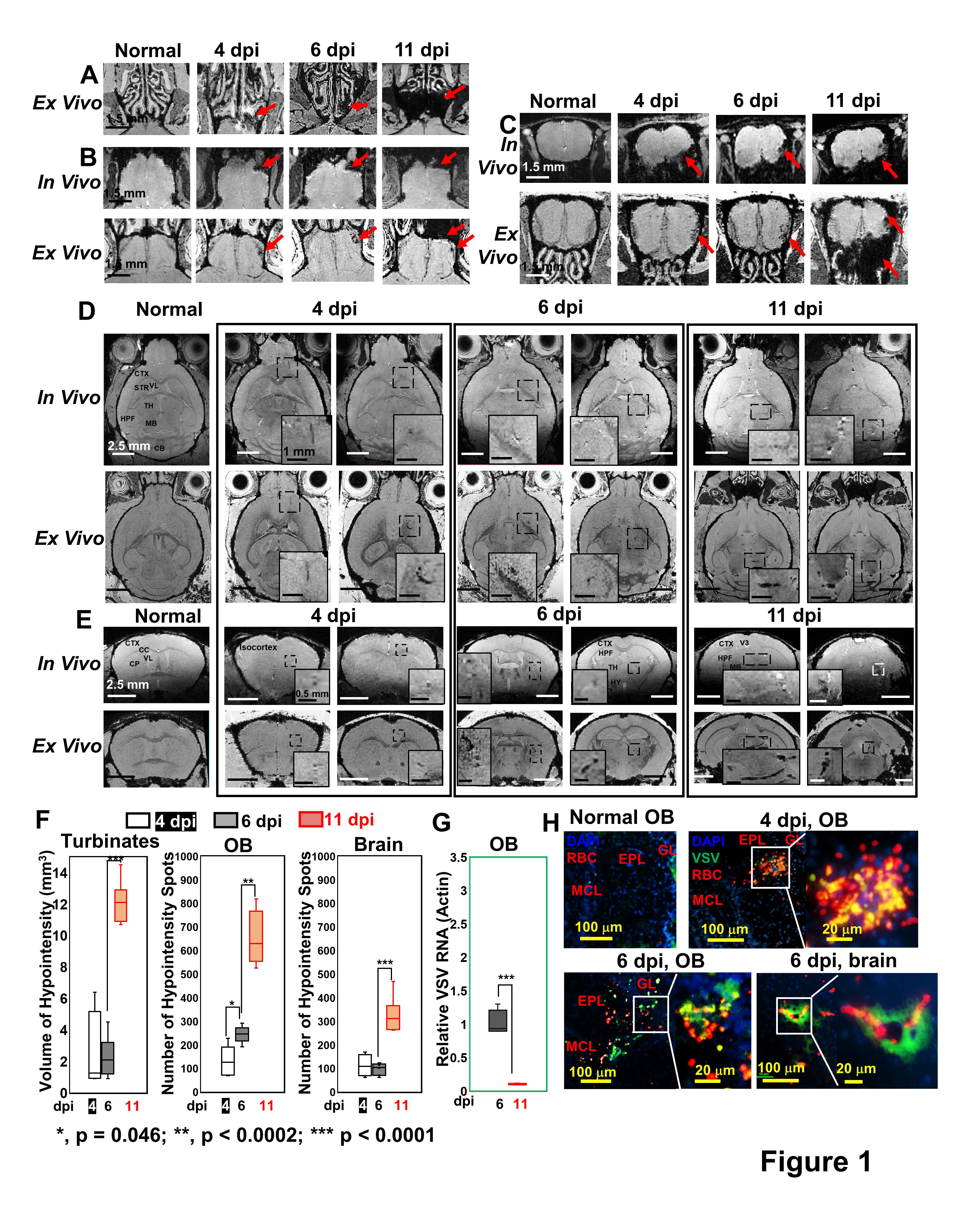
Figure 1. MRI detected microbleeds in turbinates, OB, and frontal brain since 4-dpi and monitored the
vessel breakdown during VSV brain infection. MR images of the turbinates (A),
OB (B, C), and brain (D, E) from normal mouse and VSV-infected mice on 4, 6,
and 11-dpi. (D, E) The inserted figures were the enlarged views of the
framed bleeding sites in the full views. Red arrow, bleeds. (F) Quantification of volume of hypointensity
at the turbinates and numbers of hypointensity spots at the OB and brain. (G) Viral titers at the OB on 6 and 11-dpi. (H) IHC staining of the OB and
brain.