Sonoko Oshima1, Yasutaka Fushimi1, Satoshi Nakajima1, Akihiko Sakata1, Takuya Hinoda1, Sayo Otani1, Krishna Pandu Wicaksono1, Hiroshi Tagawa1, Yang Wang1, Yuichiro Sano2, Rimika Imai2, Masahito Nambu2, Koji Fujimoto3, Hitomi Numamoto4, Kanae Kawai Miyake4, Tsuneo Saga4, and Yuji Nakamoto1
1Department of Diagnostic Radiology and Nuclear Medicine, Graduate School of Medicine, Kyoto University, Kyoto, Japan, 2Canon Medical Systems Corporation, Otawara, Japan, 3Department of Real World Data Research and Development, Graduate School of Medicine, Kyoto University, Kyoto, Japan, 4Department of Advanced Medical Imaging Research, Graduate School of Medicine, Kyoto University, Kyoto, Japan
1Department of Diagnostic Radiology and Nuclear Medicine, Graduate School of Medicine, Kyoto University, Kyoto, Japan, 2Canon Medical Systems Corporation, Otawara, Japan, 3Department of Real World Data Research and Development, Graduate School of Medicine, Kyoto University, Kyoto, Japan, 4Department of Advanced Medical Imaging Research, Graduate School of Medicine, Kyoto University, Kyoto, Japan
Neuromelanin-sensitive MRI of 1 NEX using DLR noise
reduction with denoising intensity coefficient of 1.0 and edge enhancement off
showed higher image quality than other DLR patterns. The reconstructed images provided
good diagnostic ability for Parkinson’s disease.
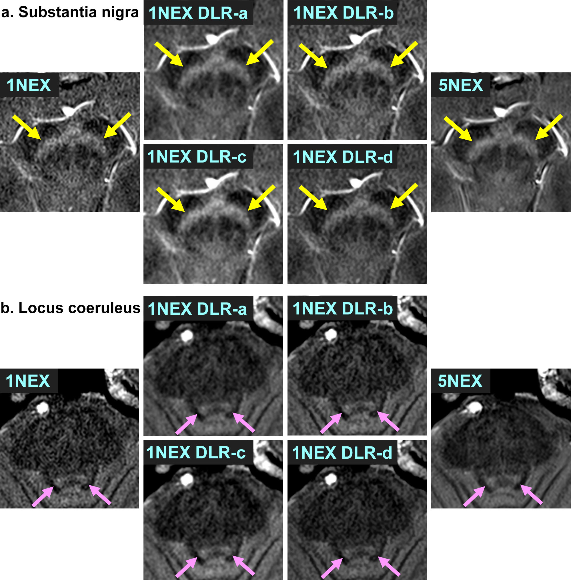
Figure 2: Images of 1 NEX without DLR, 1 NEX with DLR-a to DLR-d,
and 5 NEX of a 50-year-old healthy female. (a) the SN (yellow arrows) and (b) LC (pink arrows). DLR reduces image noise while preserving the
contrast of the SN and LC against background areas.

Figure 4: Images of the SN
(yellow arrows) and LC (pink arrows) of a 50-year-old female healthy volunteer
and a 54-year-old female patient with PD. Images of 1 NEX without DLR and 1 NEX
with DLR-c are shown. The images with DLR can visualize the SN and LC more clearly than
without DLR. The patient with PD shows decreased contrast of the SN and LC compared
to the HC.
