Félix Dumais1, Marco Perez Caceres1, Noémie Arès-Bruneau2, Christian Bocti2,3,4, and Kevin Whittingstall5
1Médecine nucléaire et radiobiologie, Université de Sherbrooke, Sherbrooke, QC, Canada, 2Faculté de Médecine et des Sciences de la Santé, Université de Sherbrooke, Sherbrooke, QC, Canada, 3Clinique de la Mémoire et Centre de Recherche sur le Vieillissement, CIUSSS de l’Estrie-CHUS, Sherbrooke, QC, Canada, 4Service de Neurologie, Département de Médecine, CHUS, Sherbrooke, QC, Canada, 5Radiologie diagnostique, Université de Sherbrooke, Sherbrooke, QC, Canada
1Médecine nucléaire et radiobiologie, Université de Sherbrooke, Sherbrooke, QC, Canada, 2Faculté de Médecine et des Sciences de la Santé, Université de Sherbrooke, Sherbrooke, QC, Canada, 3Clinique de la Mémoire et Centre de Recherche sur le Vieillissement, CIUSSS de l’Estrie-CHUS, Sherbrooke, QC, Canada, 4Service de Neurologie, Département de Médecine, CHUS, Sherbrooke, QC, Canada, 5Radiologie diagnostique, Université de Sherbrooke, Sherbrooke, QC, Canada
Neural network performances are similar to those
obtained with trained annotators on large arteries. We can do a multilabel
brain artery segmentation by propagating CW annotation through the arterial
system. The variability of this algorithm to compute diameters is smaller than
1 voxel.
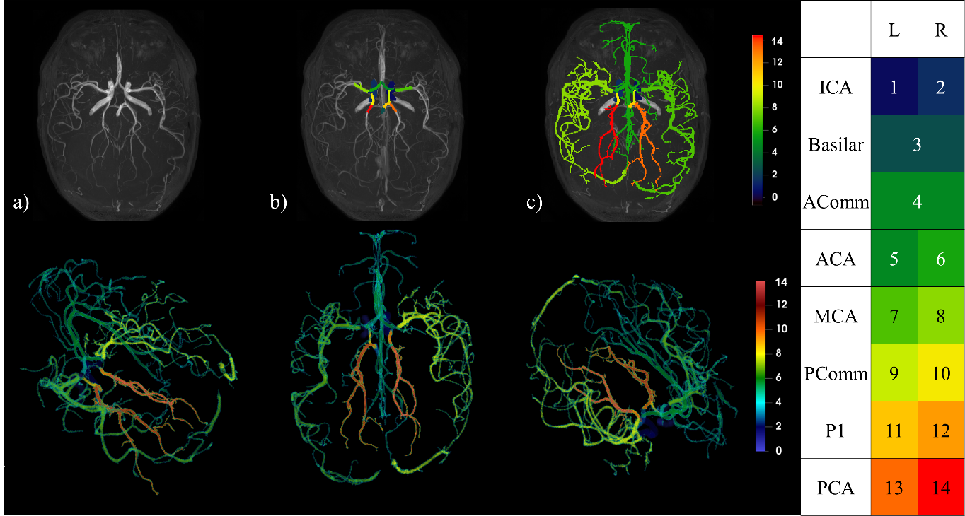
Figure 2: Top row : a) Raw TOF, b) CW segmentation,
c) Propagation of CW labels in the brain; Bottom row: 3D rendering of a full
arterial segmentation; Right: Legend indicating artery labels with their
corresponding color.
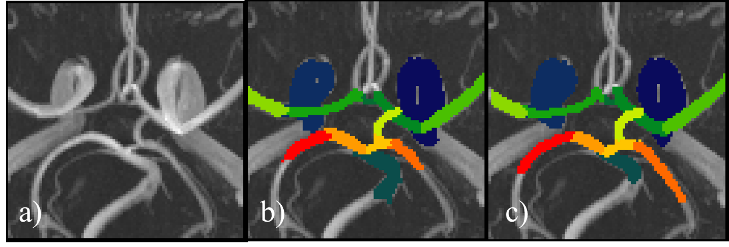
Figure 3: CW from a TOF-MRA raw (a) alongside its
manual annotation
(b) and the neural network
prediction (c)