Miaoqi Zhang1, Qingchu Jin2, Mingzhu Fu1, Hanyu Wei1, and Rui Li1
1Center for Biomedical Imaging Research, Department of Biomedical Engineering, Tsinghua University, Beijing, China, 2Johns Hopkins University, Baltimore, MD, United States
1Center for Biomedical Imaging Research, Department of Biomedical Engineering, Tsinghua University, Beijing, China, 2Johns Hopkins University, Baltimore, MD, United States
we successfully segmented IAs from dual inputs (TOF-MRA and T1-VISTA) using the hyperdense net with higher accuracy than a single input.

Figure 2. Four IA segmentation examples.
Each row represents a patient in the test set. Six columns from left to right
represent TOF-MRA, T1-VISTA, ground truth (GT), segmentation from the model
with dual inputs, segmentation from the model with TOF-MRA alone and
segmentation from the model with T1-VISTA alone.
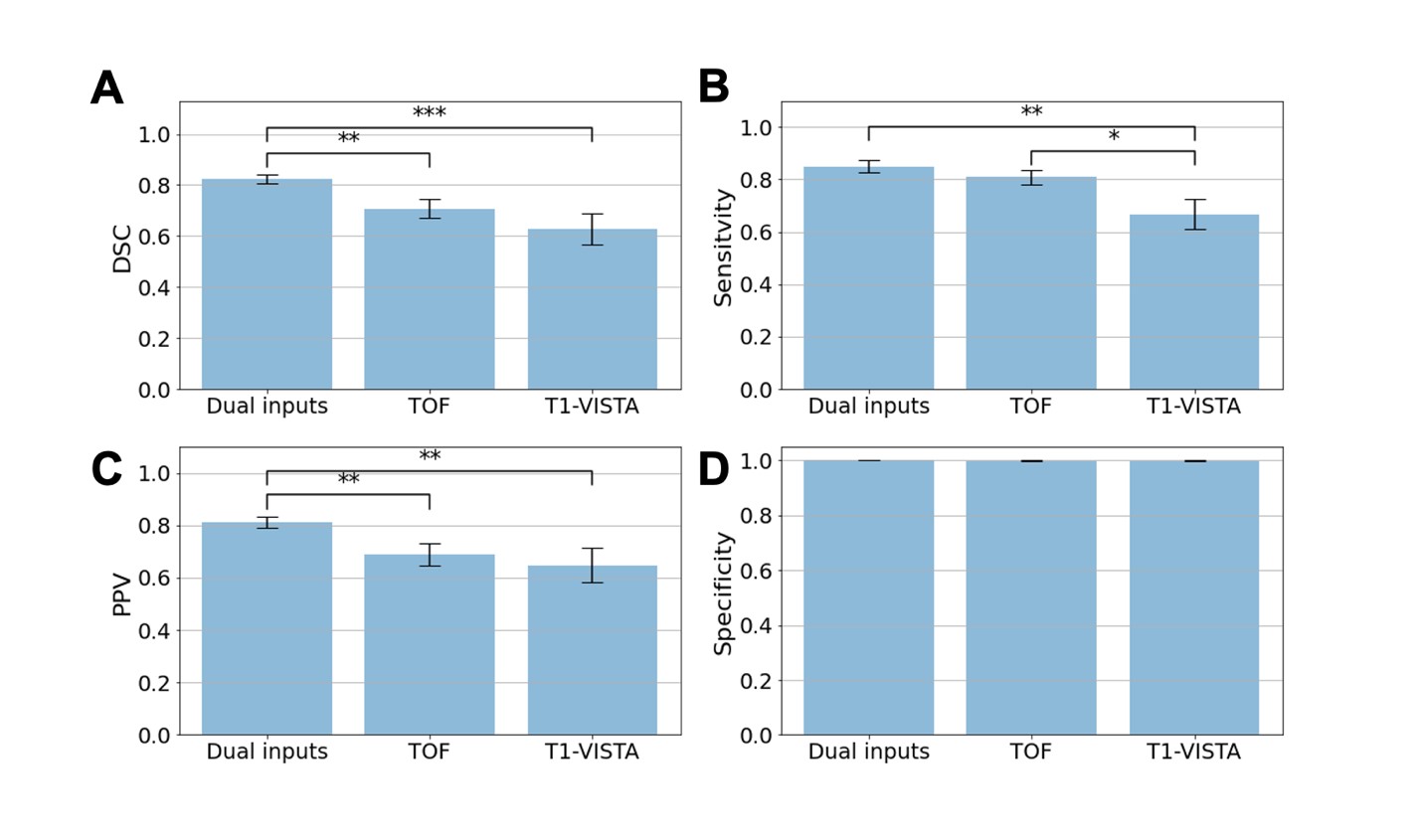
Figure 3. Aneurysm segmentation evaluation
across different combinations of image inputs: dual inputs, TOF-MRA alone and
T1-VISTA alone. (A) Sørensen–Dice coefficient (DSC); (B) sensitivity; (C) positive
predictive value (PPV); and (D) specificity. Paired Student’s t-tests were
performed with the notation *: P < 0.05; **: P < 0.005; ***: P <
0.0005.