Suvai Gunasekaran1, Julia Hwang1, Daming Shen1,2, Aggelos Katsaggelos1,3, Mohammed S.M. Elbaz1, Rod Passman4, and Daniel Kim1,2
1Radiology, Northwestern University, Feinberg School of Medicine, Chicago, IL, United States, 2Biomedical Engineering, Northwestern University, Evanston, IL, United States, 3Electrical and Computer Engineering, Northwestern University, Evanston, IL, United States, 4Cardiology, Northwestern University, Feinberg School of Medicine, Chicago, IL, United States
1Radiology, Northwestern University, Feinberg School of Medicine, Chicago, IL, United States, 2Biomedical Engineering, Northwestern University, Evanston, IL, United States, 3Electrical and Computer Engineering, Northwestern University, Evanston, IL, United States, 4Cardiology, Northwestern University, Feinberg School of Medicine, Chicago, IL, United States
A proposed 2 s automated left atrial wall segmentation from 3D
late gadolinium enhancement imaging using deep learning provided comparable segmentation
performance to 16-30 min manual segmentation.
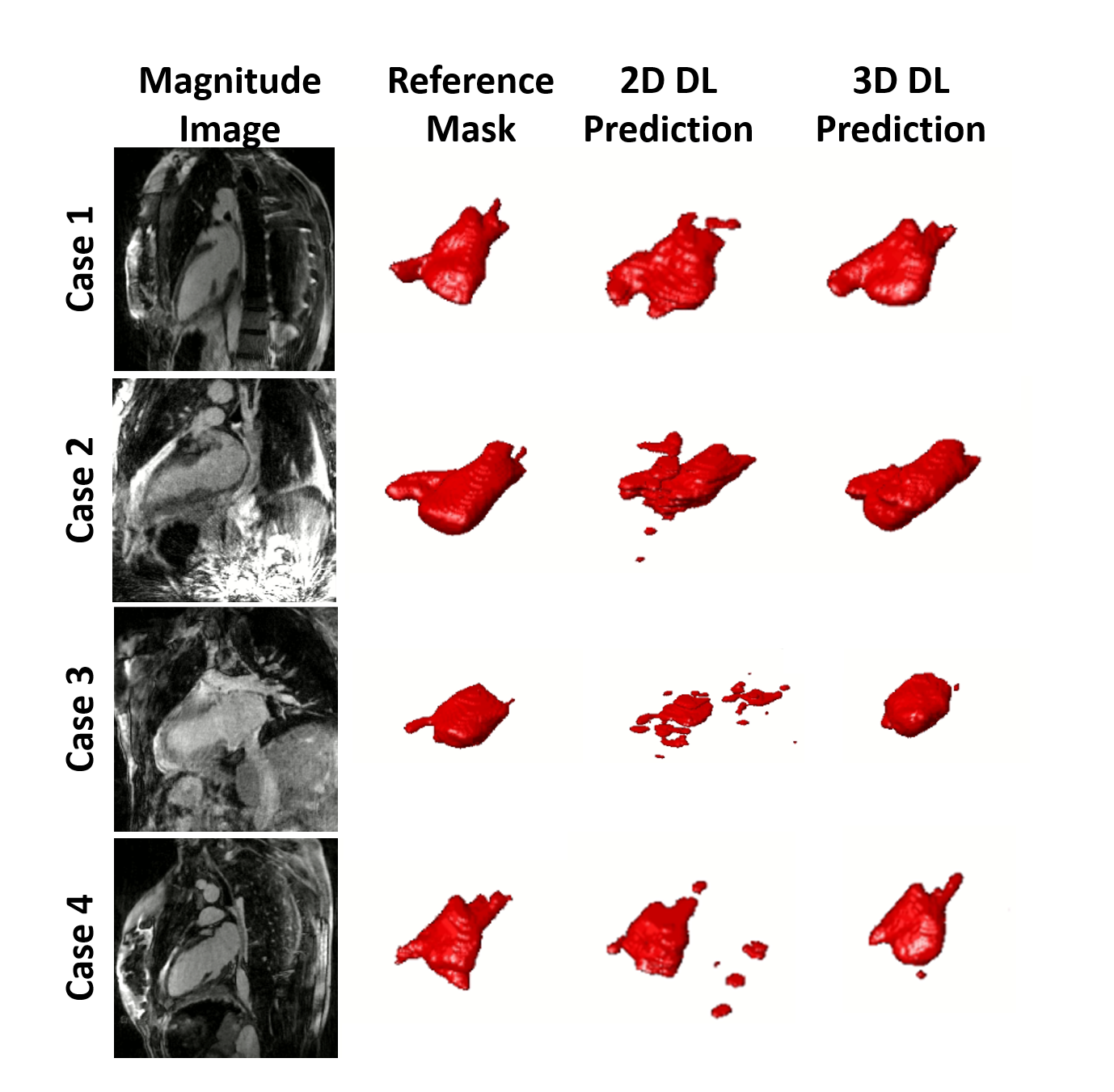
Figure 3. Examples of results for the different
DL networks from four testing cases. The segmentation results generated by the 3D
inputs are qualitatively better than those generated by the 2D inputs.
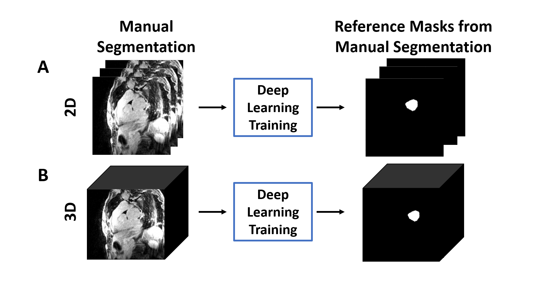
Figure 2. The overall process of DL
segmentation for (A) 2D and (B) 3D inputs. The LA LGE and reference masks
extracted from manual contours were used as input and reference to train the DL
network. For testing, the LA LGE images were fed into the trained network to
get the DL segmented masks.