Lena Vaclavu1, Dilek Betül Arslan2, Lydiane Hirschler1, Carles Falcon3,4, Esin Ozturk-Isik2, Juan Domingo Gispert3,4, Paula Montesinos5, Kim van de Ven6, and Matthias JP van Osch1
1Department of Radiology, C.J. Gorter Center for High Field MRI, Leiden University Medical Center, Leiden, Netherlands, 2Biomedical Engineering Institute, Boğaziçi University, Istanbul, Turkey, 3BarcelonaBeta Brain Research Center (BBRC), Pasqual Maragall Foundation, Barcelona, Spain, 4CIBER-BBN, Madrid, Spain, 5Philips Healthcare Iberia, Madrid, Spain, 6Philips Healthcare, Best, Netherlands
1Department of Radiology, C.J. Gorter Center for High Field MRI, Leiden University Medical Center, Leiden, Netherlands, 2Biomedical Engineering Institute, Boğaziçi University, Istanbul, Turkey, 3BarcelonaBeta Brain Research Center (BBRC), Pasqual Maragall Foundation, Barcelona, Spain, 4CIBER-BBN, Madrid, Spain, 5Philips Healthcare Iberia, Madrid, Spain, 6Philips Healthcare, Best, Netherlands
Our results show
that increased signal fluctuations in arterial vessels in time-encoded pCASL is
associated with selective background suppression and is independent of the Hadamard
labeling process.

Figure 1. a) An example of the shine-through effect in a
healthy volunteer seen in the perfusion block of a fully acquired time-encoded
pCASL (te-pCASL) dataset noticeable as negative signal in the large arteries
and/or positive signal in higher slices. b) The temporal standard deviation
(tStdv) of the same dataset shows that the effect (high tStdv in vessels) is
present in all post-label-delays (PLDs) and that tStdv is an insightful measure
of the effect.
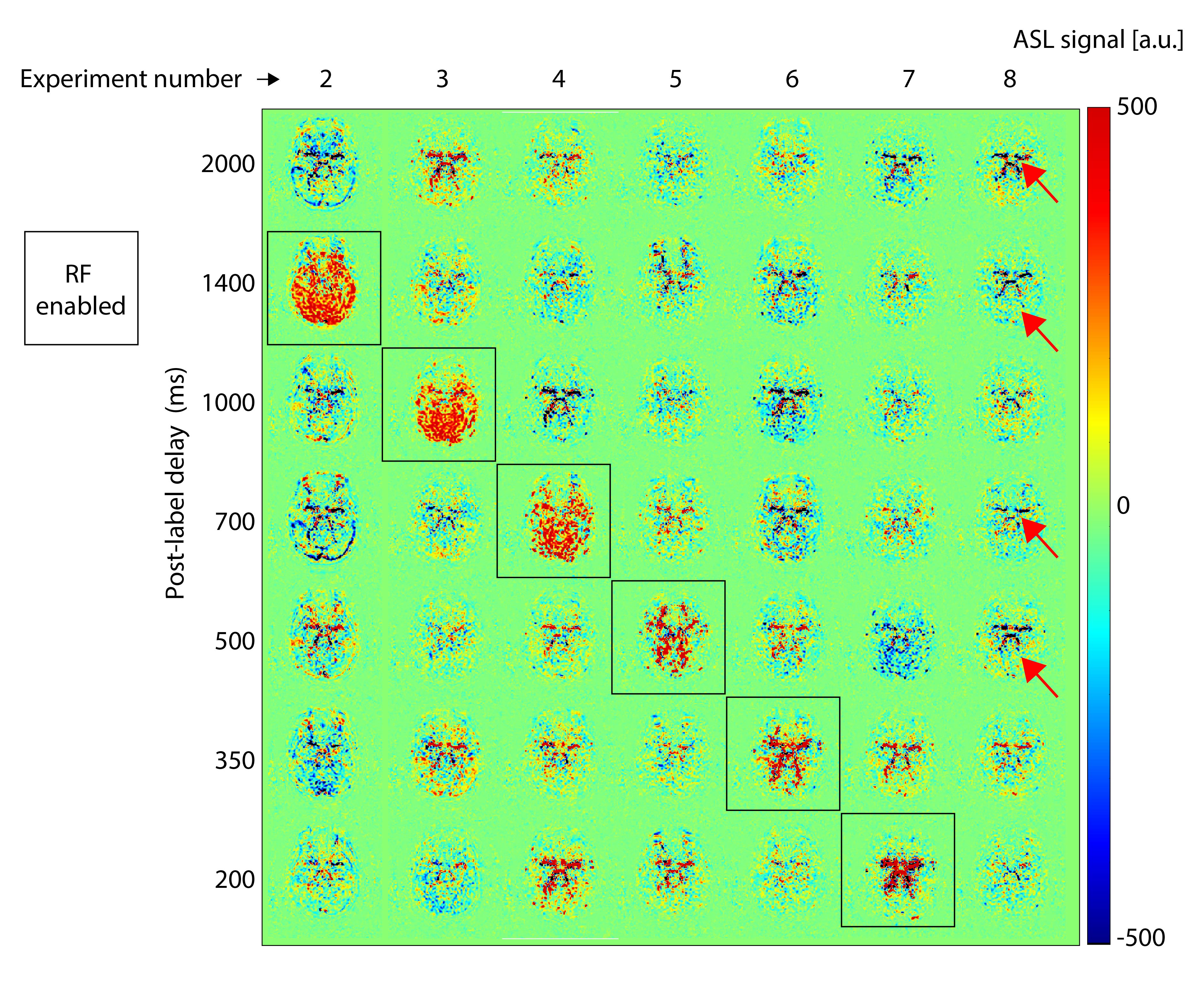
Figure 4. ASL (subtracted) signal is shown for a single
slice low in the brain, for the seven individual experiments in which RF
(labeling) was enabled for single blocks only (columns). The separation of the
individual blocks in each experiment can be seen in the black squares which
have positive ASL signal. We observed that the shine-through artefact (red
arrows) was visible in the disabled blocks, (negative or positive
signal), and surprisingly, also in the experiment without any labeling at all
(last column, experiment #8). See Figure 1 for explanation of the experiment
numbers.