Refaat E Gabr1 and Ponnada A Narayana1
1Diagnostic and Interventional Imaging, University of Texas Health Science Center at Houston, Houston, TX, United States
1Diagnostic and Interventional Imaging, University of Texas Health Science Center at Houston, Houston, TX, United States
DAWM was segmented in
MS patients using neural network. 45% of DAWM persisted and 15% converted to T2
lesions at 60 months.
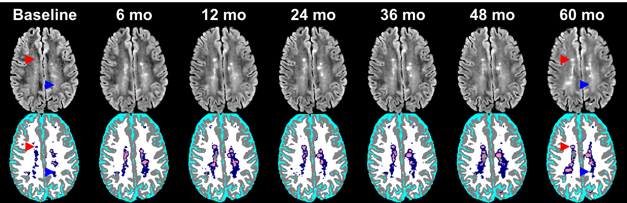
Fig. 2: FLAIR images and DL segmentation from an MS patient in the
CombiRx study over 60 months. Good delineation is obtained for brain tissue,
lesions (red), and DAWM (blue). Part of DAWM can be clearly seen to evolve into
focal lesions (red arrowhead), while other parts of DAWM (blue arrowhead)
persisted for 60 months.
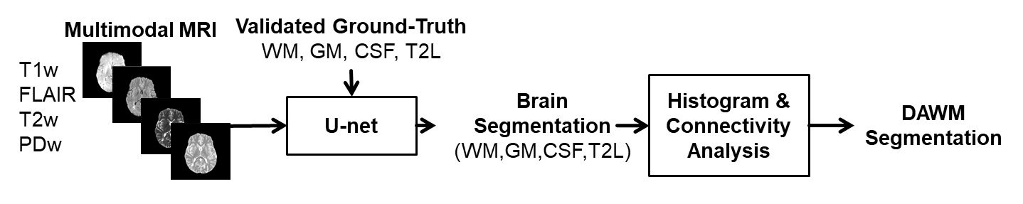
Fig. 1. DAWM
segmentation using CNN. A U-net is trained for brain segmentation
(GM, WM, CSF, T2L) from multimodal MR images. The output tissue scores are used
in histogram and connectivity analysis to obtain DAWM segmentation.