Zahra Hosseinpour1, Olayinka Oladosu2, Mahshid Soleymani1, G Bruce Pike2, and Yunyan Zhang2
1Biomedical Engineering program, Schulish School of Engineering, University of Calgary, Calgary, AB, Canada, 2Department of Radiology and Clinical Neurosciences, Hotchkiss Brain Institute, University of Calgary, Calgary, AB, Canada
1Biomedical Engineering program, Schulish School of Engineering, University of Calgary, Calgary, AB, Canada, 2Department of Radiology and Clinical Neurosciences, Hotchkiss Brain Institute, University of Calgary, Calgary, AB, Canada
Texture analysis of MRI detected several tissue types
in multiple sclerosis (MS). It identified de- and re-myelination in MS
postmortem and differentiated the highly demyelinated and potentially remyelinated
lesions in MS patients.
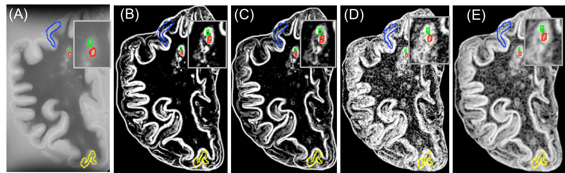
Figure 1. Postmortem T2-MRI and calculated
maps. The shown are T2 (A), T2-Contrast (B), T2-Dissimilarity (C), T2-Entropy
(D): all from average GLCM, and T2-entropy filter (E). Different colors
demonstrate different lesion types: red: WML, green: remyelinated WML, blue:
type IV cortical lesion, yellow: type III cortical lesion.
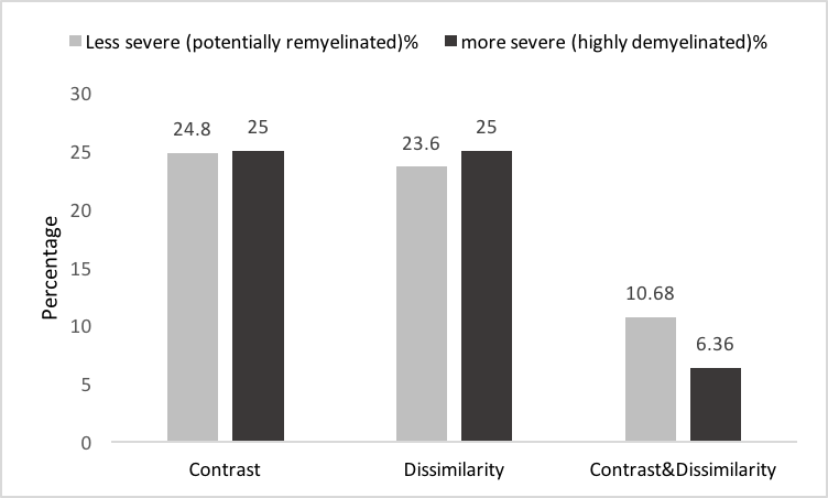
Figure 4) In-vivo lesion severity. 25th
percentile of contrast and both contrast and dissimilarity were used to detect
the less severe (potentially remyelinated) lesions. And 75th
percentile of the same parameters were applied to have more severe lesions (highly
demyelinated and axonal damaged). We found that most of the patients have both
types of lesions and the percentage of the less severe lesions is in the range
of reported remyelination percentage in MS.