Lucas Soustelle1,2, Andreea Hertanu1,2, Arnaud Le Troter1,2, Soraya Gherib1,2, Samira Mchinda1,2, Patrick Viout1,2, Lauriane Pini1,2, Claire Costes1,2, Sylviane Confort-Gouny1,2, Adil Maarouf1,2,3, Bertrand Audoin1,2,3, Audrey Rico1,2,3, Clémence Boutière1,2,3, Maxime Guye1,2, Jean-Philippe Ranjeva1,2, Gopal Varma4, David C. Alsop4, Jean Pelletier1,2,3, Olivier M. Girard1,2, and Guillaume Duhamel1,2
1Aix Marseille Univ, CNRS, CRMBM, Marseille, France, 2APHM, Hôpital Universitaire Timone, CEMEREM, Marseille, France, 3APHM, Hôpital Universitaire Timone, Service de neurologie, Marseille, France, 4Division of MR Research, Radiology, Beth Israel Deaconess Medical Center, Harvard Medical School, Boston, MA, United States
1Aix Marseille Univ, CNRS, CRMBM, Marseille, France, 2APHM, Hôpital Universitaire Timone, CEMEREM, Marseille, France, 3APHM, Hôpital Universitaire Timone, Service de neurologie, Marseille, France, 4Division of MR Research, Radiology, Beth Israel Deaconess Medical Center, Harvard Medical School, Boston, MA, United States
Active
MS lesions were evaluated over a 12-month follow-up study by modelling the
signal evolution of ihMT, conventional MT, DTI and T1w data. Results show that MR
metrics in lesions recover at different rates, emphasizing different sensitivity
to MS physiopathology mechanisms.
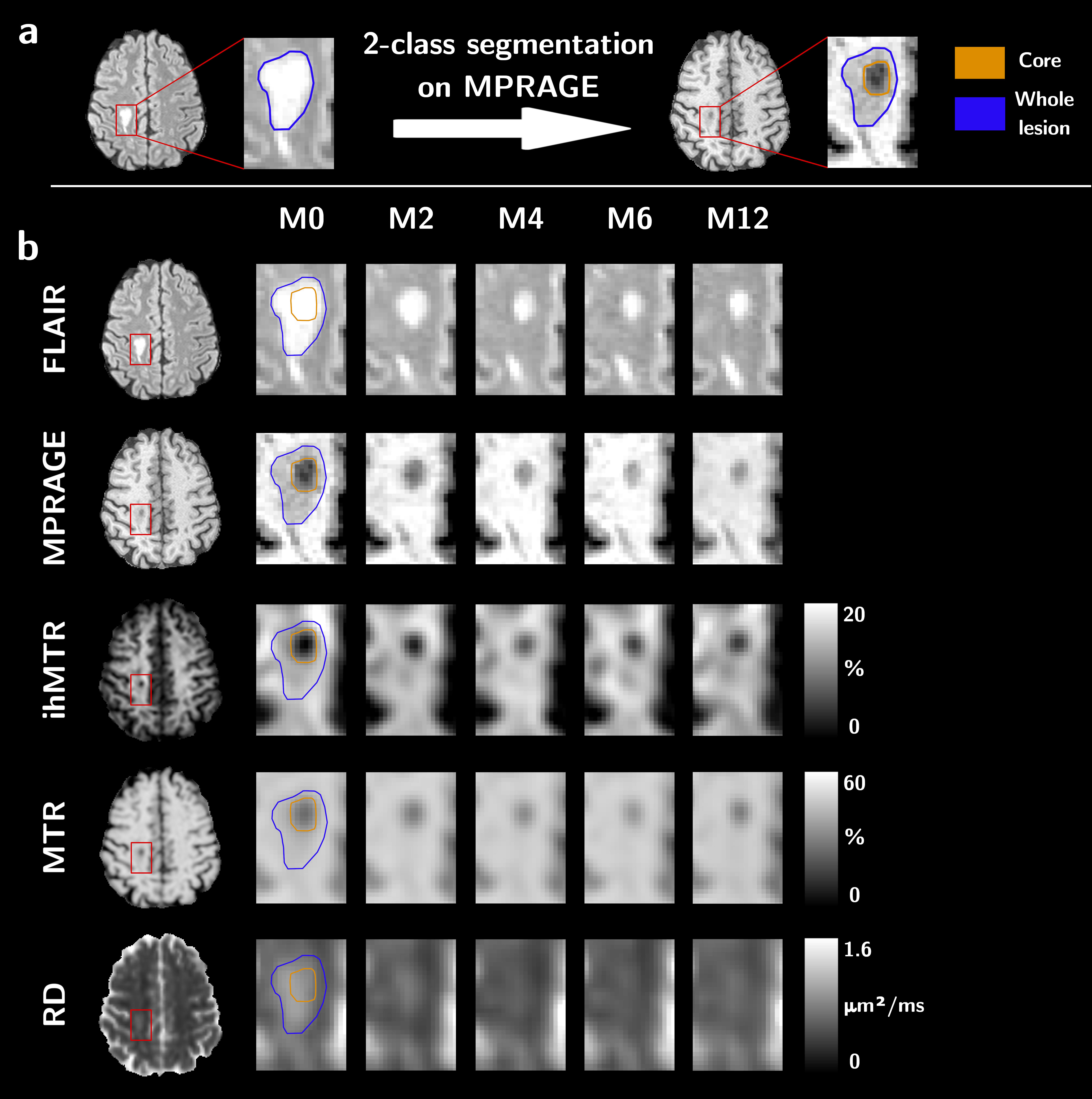
Figure 1: Sketch of the sub-segmentation processing of the lesion into core
and edge regions based on the T1w contrast and from the manually drawn mask on
the FLAIR image (a), and illustrative depiction of the evolution of MR
contrasts over time in an active lesion (b).

Figure 3: Example of an RVtoC curve adjustment over an ihMTR lesion in the
core region (a), and boxplots of the average recovery rates per patients of
ihMTR, MTR, RD and MPRAGE (b). Note that patient P01 was not included as the
dynamic of its two lesions could not be resolved by the fitting model for both
ihMTR and RD (ρ²<0.75).
