Tomoko Maekawa1,2, Akifumi Hagiwara1,3, Masaaki Hori1,4, Christina Andica1, Shohei Fujita1,5, Toshiaki Akashi1, Koji Kamagata1, Akihiko Wada1, and Shigeki Aoki1
1Department of Radiology, Juntendo University School of Medicine, Tokyo, Japan, 2Division of Regenerative Medicine, The Jikei University School of Medicine, Tokyo, Japan, 3Department of Radiological Science, David Geffen School of Medicine, University of California Los Angeles, Los Angeles, CA, United States, 4Department of Diagnostic Radiology, Toho University Omori Medical Center, Tokyo, Japan, 5Department of Radiology, Graduate School of Medicine, The University of Tokyo, Tokyo, Japan
1Department of Radiology, Juntendo University School of Medicine, Tokyo, Japan, 2Division of Regenerative Medicine, The Jikei University School of Medicine, Tokyo, Japan, 3Department of Radiological Science, David Geffen School of Medicine, University of California Los Angeles, Los Angeles, CA, United States, 4Department of Diagnostic Radiology, Toho University Omori Medical Center, Tokyo, Japan, 5Department of Radiology, Graduate School of Medicine, The University of Tokyo, Tokyo, Japan
We investigated the association between estimated time from the onset of multiple sclerosis plaques and myelin- and axon-related quantitative MRI measurements. Multiple sclerosis plaques with longer estimated time from the onset had significantly lower myelin volume fraction.
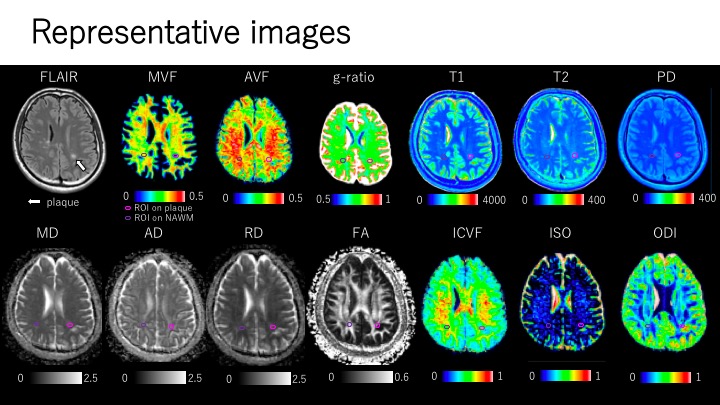
Figure 3. Representative quantitative maps of an MS patient. Plaques were defined as a white matter area of more than 5 mm in diameter, with abnormally high intensity on synthetic FLAIR images. Each plaque was manually segmented. All the ROIs placed on synthetic FLAIR images were copied and pasted onto the quantitative maps.
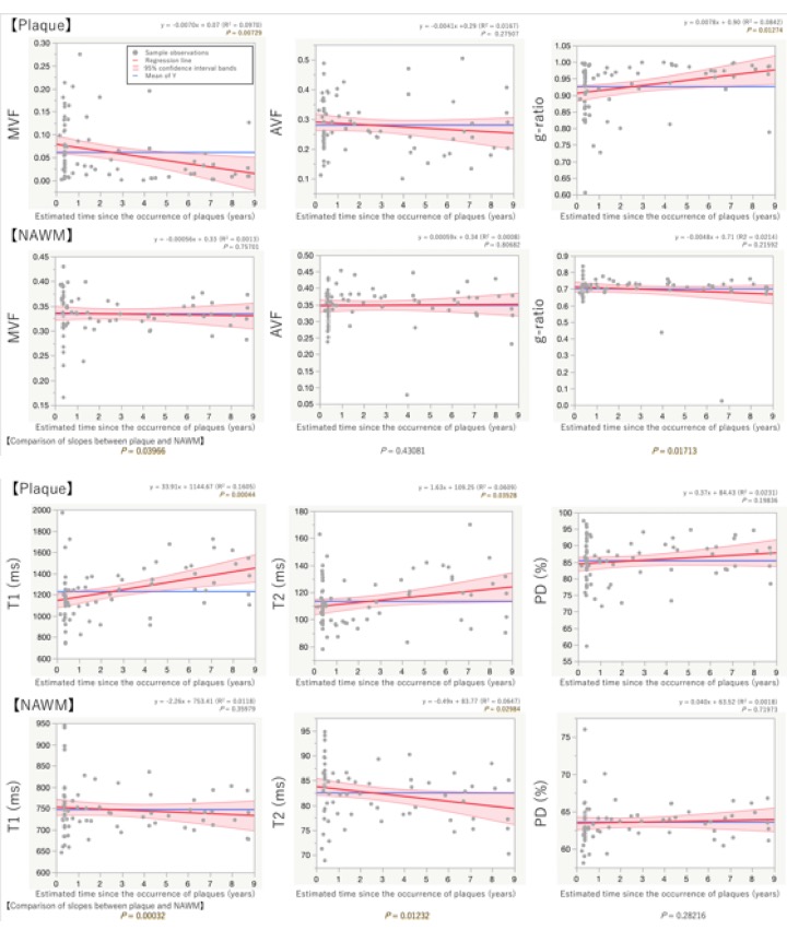
Figure 4. Simple linear regressions to assess the association between estimated time from the onset of plaques and quantitative MRI metrics. Plaque MVF and g-ratio in relation to the estimated time from the onset were significantly fitted to linear lines with negative and positive slopes, respectively. Plaque T1 and T2 in relation to the estimated time from the onset were significantly fitted to linear lines with positive slopes. NAWM T2 in relation to the estimated time from the onset was significantly fitted to a linear lineswith a negative slope.