Rina Ito1,2, Yuji Komaki2, Fumiko Seki2, Mayu Iida1,2, Mitsuki Rikitake1,2, Marin Nishio1,2, Junichi Hata1,3, and Takako Shirakawa1
1Department of Radiological Sciences, Human Health Sciences, Tokyo Metropolitan University, Tokyo, Japan, 2Live imaging Center, Central Institute for Experimental Animals, Kanagawa, Japan, 3Jikei University Graduate School of Medicine, Tokyo, Japan
1Department of Radiological Sciences, Human Health Sciences, Tokyo Metropolitan University, Tokyo, Japan, 2Live imaging Center, Central Institute for Experimental Animals, Kanagawa, Japan, 3Jikei University Graduate School of Medicine, Tokyo, Japan
The
number of resting-state networks and its strength increased dramatically with
age until common marmosets become adults (the age of 24 months) and declined
steadily at older age after 24 months.
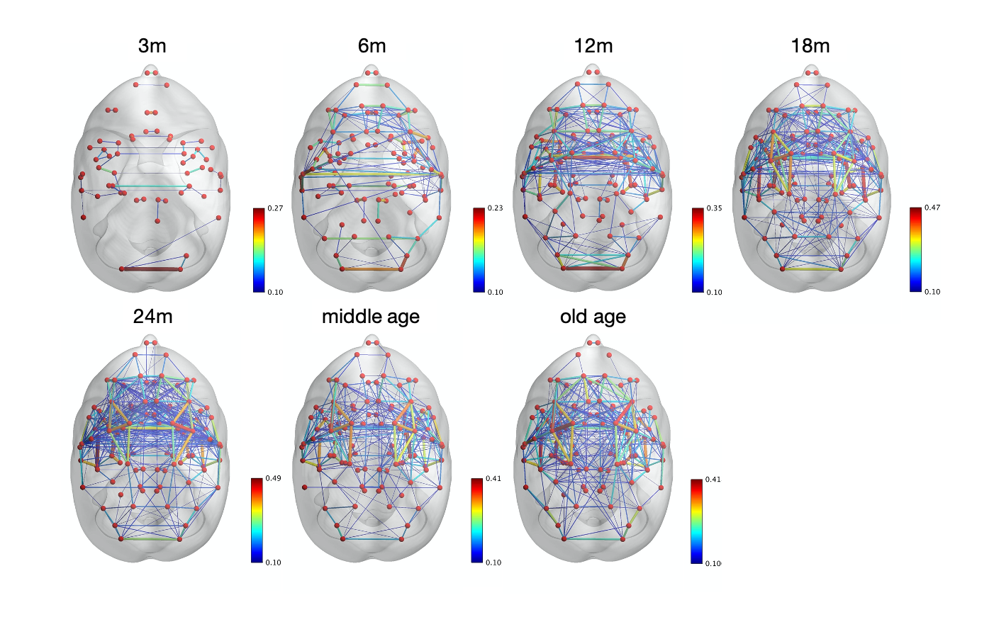
Figure 1. Images of functional brain networks
by developmental stage
The red points “node” represents anatomical
regions, and the lines “edge” shows measurement of functional connectivity (FC)
between pairs of nodes. The number of nodes and edges was getting larger with
age. The scale bar was put on right side. Binary threshold was set 0.1 in all
months of age.
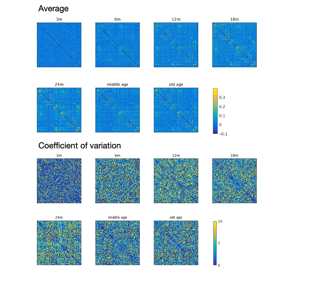
Figure 2. FC matrix by developmental stage
The brain was parcellated into 51 labels, and
FC matrix was composed by the connection of each labels. The scale bar on the
right side shows the strength of positive and negative connectivity. The upper
and lower images represent averaged and coefficient of variation FC matrix for
each. The development of FC and large variation were indicated as marmosets
grow.