Yongxian Qian1, Gregory Chang1, Eric J. Strauss2, and Fernando E. Boada1
1Radiology, New York University, New York, NY, United States, 2Orthopaedic Surgery, New York University, New York, NY, United States
1Radiology, New York University, New York, NY, United States, 2Orthopaedic Surgery, New York University, New York, NY, United States
This human study at 7T presents the great potential of ultra-high
resolution UTE imaging to simultaneously visualize cartilage, meniscus,
ligament, tendon, and even the chondro-osseous junction in the knee joint, critical
to understanding the onset and progression of osteoarthritis.
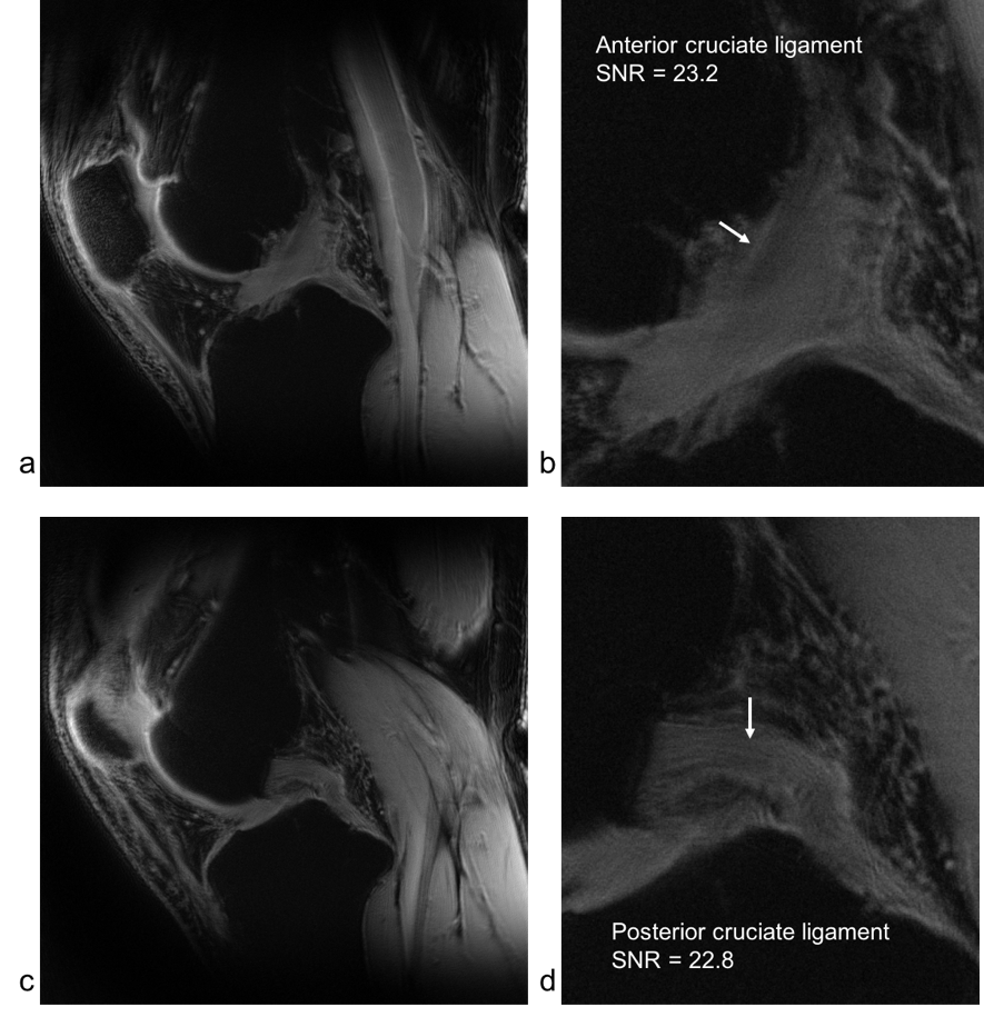
Fig. 5.
Anterior and posterior cruciate ligaments (ACL and PCL) in the ultra-high
resolution UTE image at 7T for the same subject as in Fig. 1: a) full
FOV and b)
local
view for ACL, and c) full FOV and d) local view for PCL.
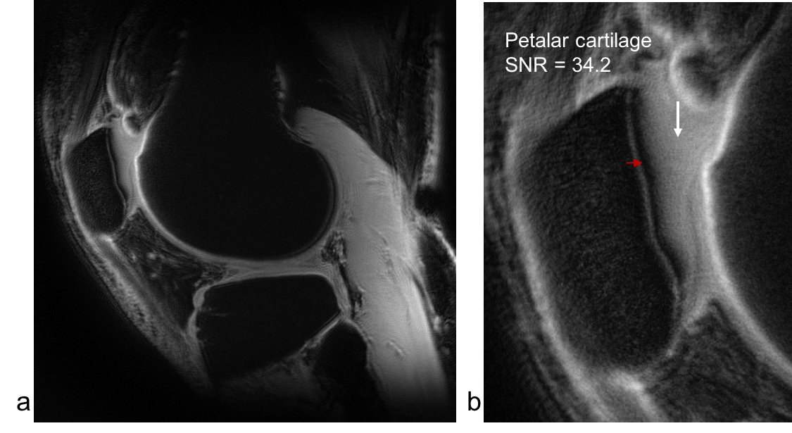
Fig. 2.
Patellar cartilage
(long white arrow) and the chondro-osseous
junction
(red arrow, black region) in the ultra high resolution UTE image at 7T
for
the same subject as in Fig. 1: a) full
FOV, and b)
local
view.
A suspicious defect is clearly visible on
the
chondro-osseous
junction above the red arrow in b).