Yuxi Pang1, Riann Palmieri-Smith2,3, and Tristan Maerz3
1Dept. of Radiology, University of Michigan, Ann Arbor, MI, United States, 2School of Kinesiology, University of Michigan, Ann Arbor, MI, United States, 3Dept. of Orthopaedic Surgery, University of Michigan, Ann Arbor, MI, United States
1Dept. of Radiology, University of Michigan, Ann Arbor, MI, United States, 2School of Kinesiology, University of Michigan, Ann Arbor, MI, United States, 3Dept. of Orthopaedic Surgery, University of Michigan, Ann Arbor, MI, United States
An efficient quantitative $$$R_{1\rho}$$$ dispersion MR imaging protocol has been
developed for clinical studies of human knee cartilage at 3T, by simultaneously tailoring spin-lock duration and strength.
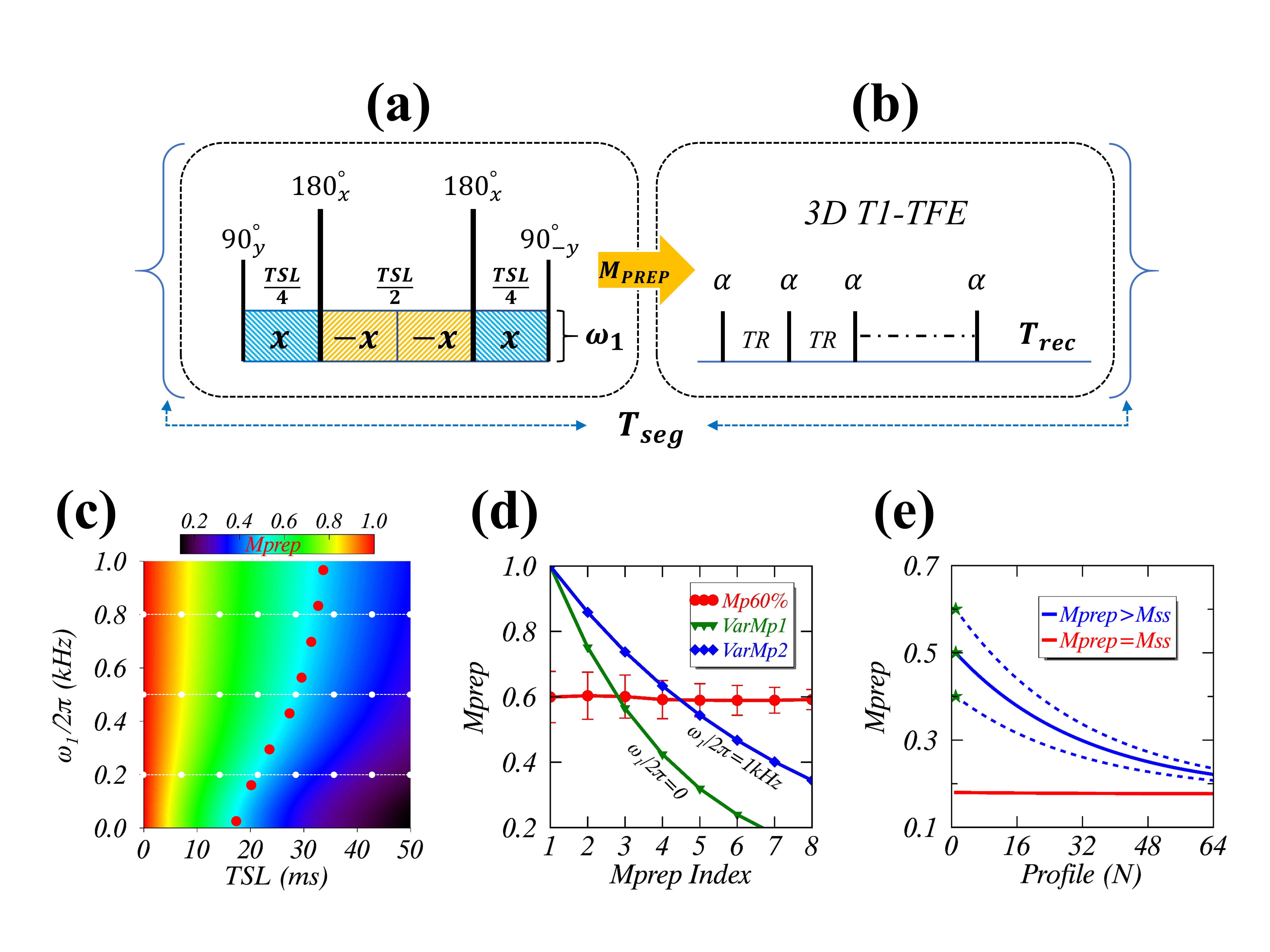
FIGURE 1. The proposed $$$R_{1\rho}$$$ dispersion imaging method including a new SL scheme (a) for turbo-FLASH (b), and a prepared constant $$$M_{prep}$$$ (red dots), with respect to the varying ones (white dots) in the standard acquisition approach (c). The $$$M_{prep}$$$ dynamic range differs significantly between from the proposed (red) and from the standard (green and blue) methods (d), whereas a cluster (blue lines) of $$$M_{prep}$$$ evolve similarly toward steady-state $$$M_{SS}$$$ (red line) during FLASH imaging readout (e).
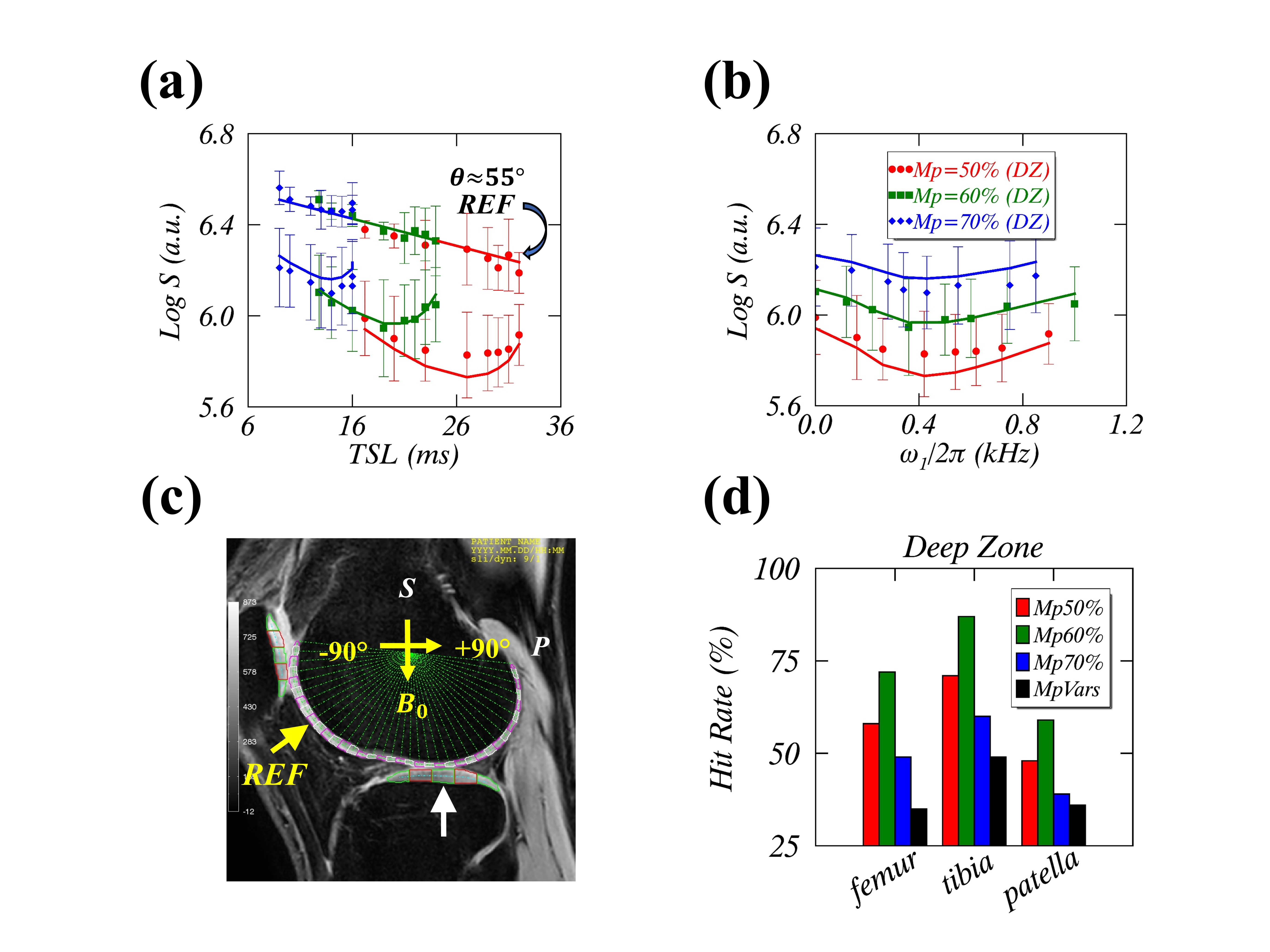
FIGURE 2. The
measured (symbols) and modeled (lines) $$$R_{1\rho}$$$ dispersion profiles with three protocols (a-b), i.e. $$$M_{prep}$$$=50% (red), 60% (green), 70% (blue). The
presented $$$R_{1\rho}$$$-weighted signals and REF datasets were taken from the segmented ROIs in the deep tibial (white arrow) and
femoral cartilage (yellow arrow) (c).
The differences between success fitting (or hit) rates (%) for modeling $$$R_{1\rho}$$$ dispersion in the deep cartilage using the
current ($$$M_{prep}$$$=50-70%) and the previous ($$$MpVars$$$) protocols (d).