Qi Zhao1, Rees P. Ridout1, Jikai Shen1, and Nian Wang2
1Duke University, Durham, NC, United States, 2Radiology and Imaging Sciences, Indiana University, Indianapolis, IN, United States
1Duke University, Durham, NC, United States, 2Radiology and Imaging Sciences, Indiana University, Indianapolis, IN, United States
The tract length and volume are sensitive to both the b value and angular resolution. In order to obtain consistent DTI outputs and tractography, the scan may require a proper b value (ranging from 500 to 1500 s/mm2) and sufficient angular resolution (14 or higher) with SNR>10.
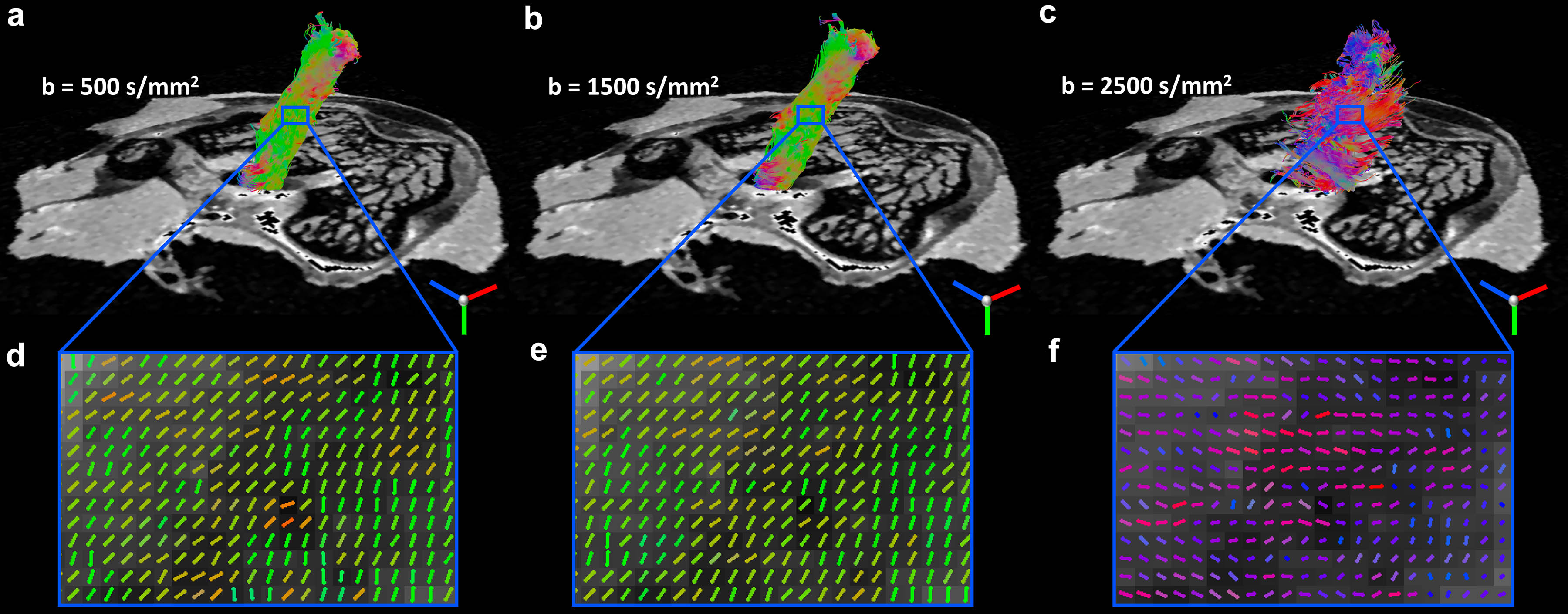
Figure 5. Diffusion tractography of ligament at different b value. The tracts (a, b) and collagen fiber directions (d, e) were visually comparable between b value of 500 s/mm2 and 1500 s/mm2. Both fiber direction estimation and tractography failed at b value of 2500 s/mm2 (c, f).
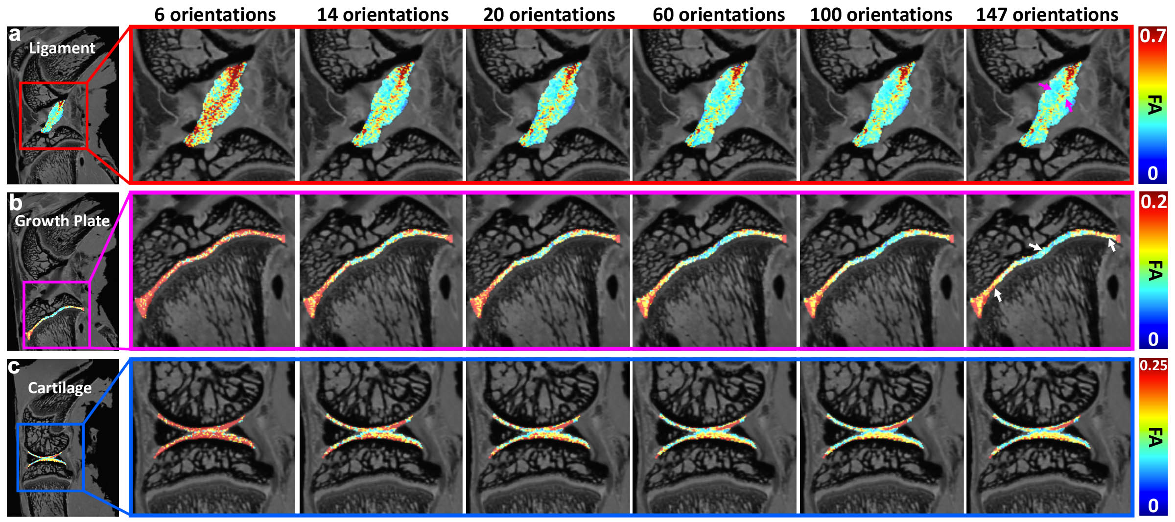
Figure 1. The FA maps of ligament, growth plate, and articular cartilage at different angular resolution. The FA values were largely overestimated with the angular resolution of 6. The values showed visually little differences when the angular resolution is higher than 14. Higher FA values were found in the center part of the ligament (purple arrows). Lower values were found in the center part of growth plate (white arrows).