Azadeh Sharafi1, Marcelo V. W. Zibetti1, Gregory Chang1, Martijn Cloos2, and Ravinder Regatte1
1Radiology, NYU Langone Health, New York, NY, United States, 2University of Queensland, Brisbane, Australia
1Radiology, NYU Langone Health, New York, NY, United States, 2University of Queensland, Brisbane, Australia
Bilateral T1, T2,
and T1ρ relaxation times, and B1+ maps can be
acquired simultaneously from hip
joints using the proposed MRF sequence.

Figure 2. Representative bilateral PD, T1, T2,
T1ρ, and B1+ maps of the hip articular cartilages.
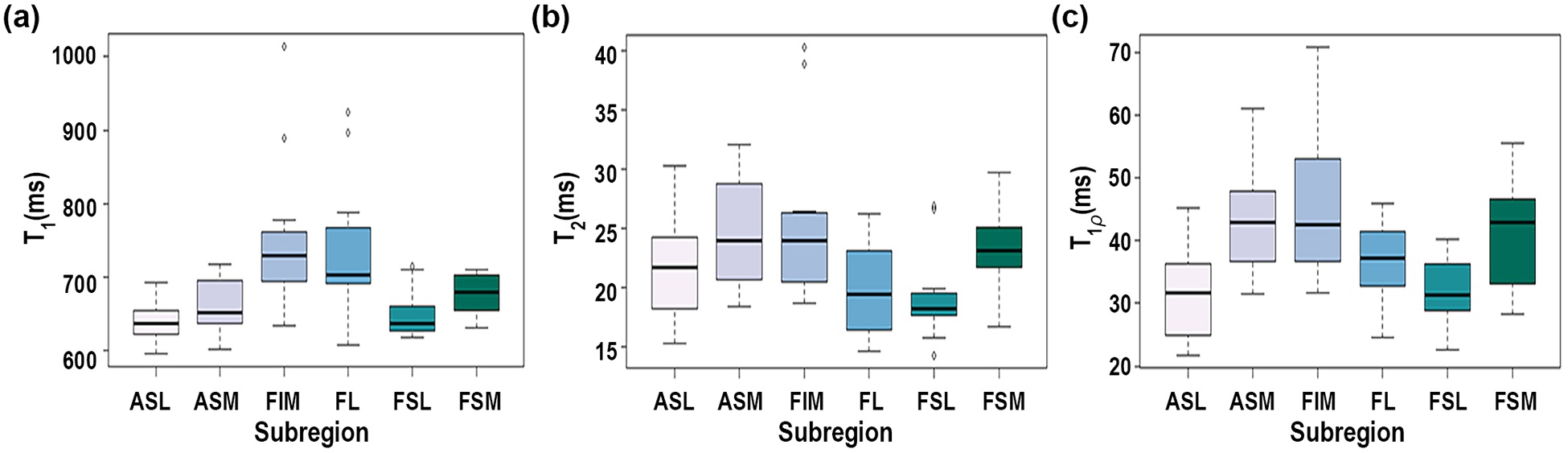
Figure 3. Boxplot comparison
between subregions for (a) T1, (b) T2, and (c) T1ρ relaxation times.