Jing jing Wu1, Xiao jun Guan2, Tao Guo2, Cheng Zhou2, Ting Gao3, Xue qin Bai2, Xiao cao Liu3, Lu yan Gu3, Pei yu Huang3, Xiao jun Xu3, and Min ming Zhang2
1Department of Radiology, The Second Affiliated Hospital, Zhejiang University School of Medicine, HangZhou, China, 2Department of Radiology, The Second Affiliated Hospital, Zhejiang University School of Medicine, Hangzhou, China, 3The Second Affiliated Hospital, Zhejiang University School of Medicine, Hangzhou, China
1Department of Radiology, The Second Affiliated Hospital, Zhejiang University School of Medicine, HangZhou, China, 2Department of Radiology, The Second Affiliated Hospital, Zhejiang University School of Medicine, Hangzhou, China, 3The Second Affiliated Hospital, Zhejiang University School of Medicine, Hangzhou, China
Two variants, rs602201 and rs198440, were found to have a positive impact on nigral iron deposition in PD. Specifically,
patients with rs602201 polymorphism are particularly vulnerable to iron
deposition in SN.

Figure 1 Data processing procedures.
A. The labels of subcortical nuclei in processed QSM images.
B. The framework of quality control steps.
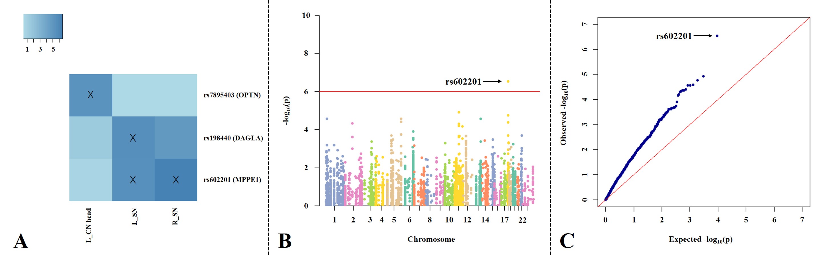
Figure 2 The association between genetic
variations and imaging phenotypes.
A. Heat map with significant associations between SNP and QT at p<10-5
(blocks labeled with “X”). Color key on the top left coded the magnitude of
-log10(p-values).
B, C. Manhattan and Q-Q plot of the most
significant association (p<10-6) (rs602201-R_SN). The horizontal
line displayed the cutoff for p<10-6. Shown on (C) is the Q-Q
plot of the distribution of the observed p-values (-log10(observed
p-value)) versus the expected p-values (-log10(expected p-value))
under the null hypothesis of no association.