Po-Yuan Chen1, Chih-Chien Tsai1, Chin-Song Lu2, Yi-Hsin Weng2, Yi-Ming Wu3, and Jiun-Jie Wang1
1Chang Gung University, Taoyuan, Taiwan, 2Chang Gung Memorial Hospital, Taoyuan, Taoyuan, Taiwan, 3Chang Gung Memorial Hospital, Linkou, Taoyuan, Taiwan
1Chang Gung University, Taoyuan, Taiwan, 2Chang Gung Memorial Hospital, Taoyuan, Taoyuan, Taiwan, 3Chang Gung Memorial Hospital, Linkou, Taoyuan, Taiwan
This study provides evidence for the
subtype-specific white matter differences in patients with MSA. Importantly,
early diagnosis for parkinsonian or cerebellar subtype of MSA is possible with
the white matter pattern by fixel-based analysis.

Figure 2. Fixels with significant (p < 0.05,
FWE-corrected) decrease in fixel-based metrics. (a)
MSA-P versus controls. Subtle changes were observed in the cohorts with disease
duration≦3 years; on the contrary, FC and FDC were
significantly decreased in the (b) MSA-C versus controls. Streamlines were
colored by direction (anterior-posterior: green; superior-inferior: blue;
left-right: red).
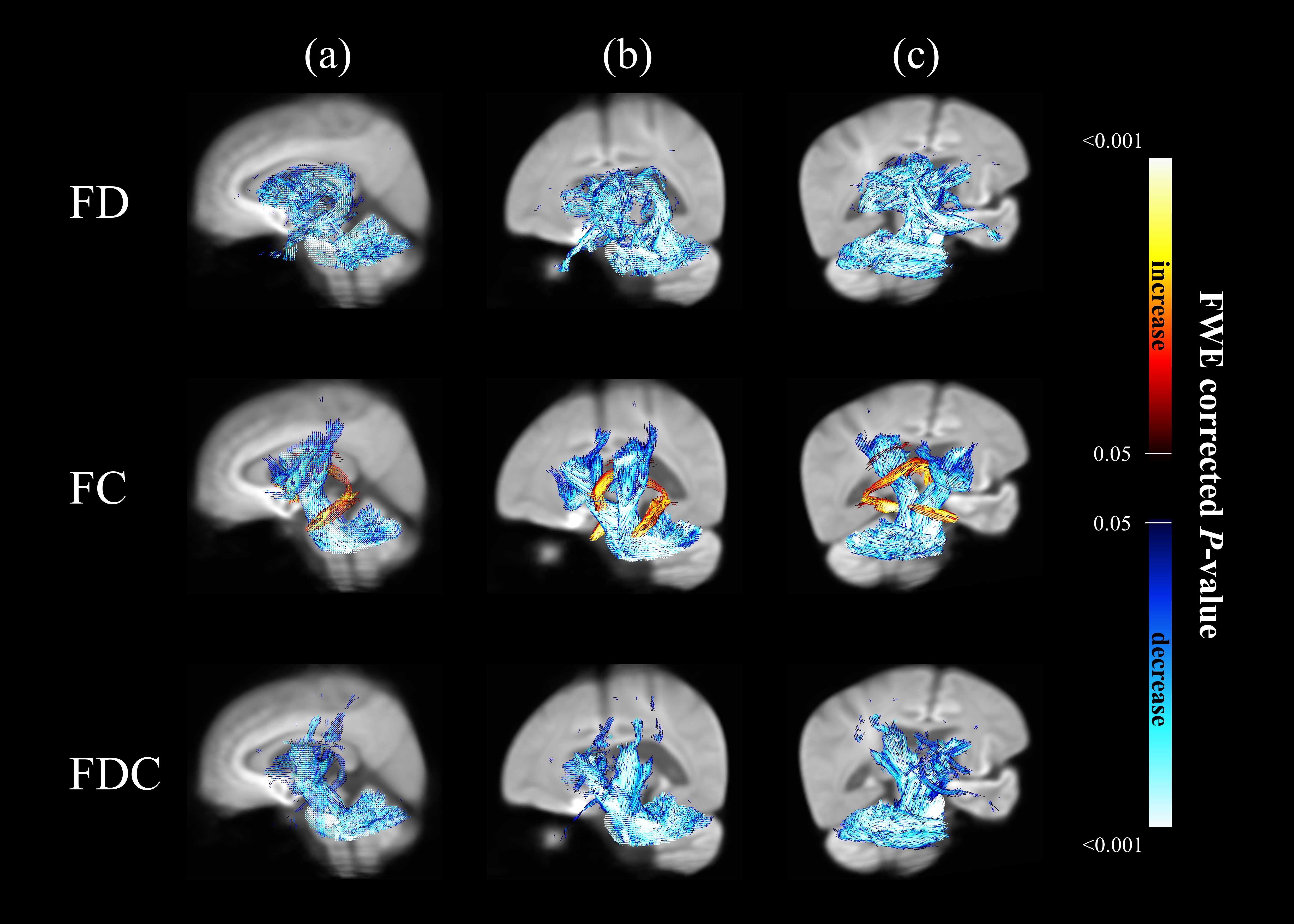
Figure 1. Significant changes in fixel-based metrics
in patients with MSA compared to healthy control subjects. Regions of significant changes in FD, FC and FDC were
displayed stereoscopically in the (a) sagittal, (b) superior left frontal and
(c) inferior right occipital view. Streamlines corresponding to significant
fixels (family-wise error corrected p <
0.05) were illustrated and colored according to p values. Cold color represented reduction, whereas warm color
represented increase.