Yao-Chia Shih1,2, Bénédicte Maréchal3,4,5, Ricardo Corredor Jerez3,4,5, Septian Hartono2,6, Hui-Hua Li2,7, Isabel Hui Min Chew1, Eng King Tan2,6, and Ling Ling Chan1,2
1Department of Diagnostic Radiology, Singapore General Hospital, Singapore, Singapore, 2Duke-NUS Medical School, Singapore, Singapore, 3Advanced Clinical Imaging Technology, Siemens Healthcare AG, Lausanne, Switzerland, 4Department of Radiology, Lausanne University Hospital and University of Lausanne, Lausanne, Switzerland, 5LTS5, École Polytechnique Fédérale de Lausanne, Lausanne, Switzerland, 6Department of Neurology, National Neuroscience Institute (Outram-campus), Singapore, Singapore, 7Health Services Research Unit, Singapore General Hospital, Singapore, Singapore
1Department of Diagnostic Radiology, Singapore General Hospital, Singapore, Singapore, 2Duke-NUS Medical School, Singapore, Singapore, 3Advanced Clinical Imaging Technology, Siemens Healthcare AG, Lausanne, Switzerland, 4Department of Radiology, Lausanne University Hospital and University of Lausanne, Lausanne, Switzerland, 5LTS5, École Polytechnique Fédérale de Lausanne, Lausanne, Switzerland, 6Department of Neurology, National Neuroscience Institute (Outram-campus), Singapore, Singapore, 7Health Services Research Unit, Singapore General Hospital, Singapore, Singapore
The automated MRPI using the proposed algorithm was expeditious (averaging
25s ± 2s per case) and reliable (average coefficient of variance <18%) as measured
on >2000 brain scans across different 1.5T and 3T MRI systems, subject cohorts
and time-points.
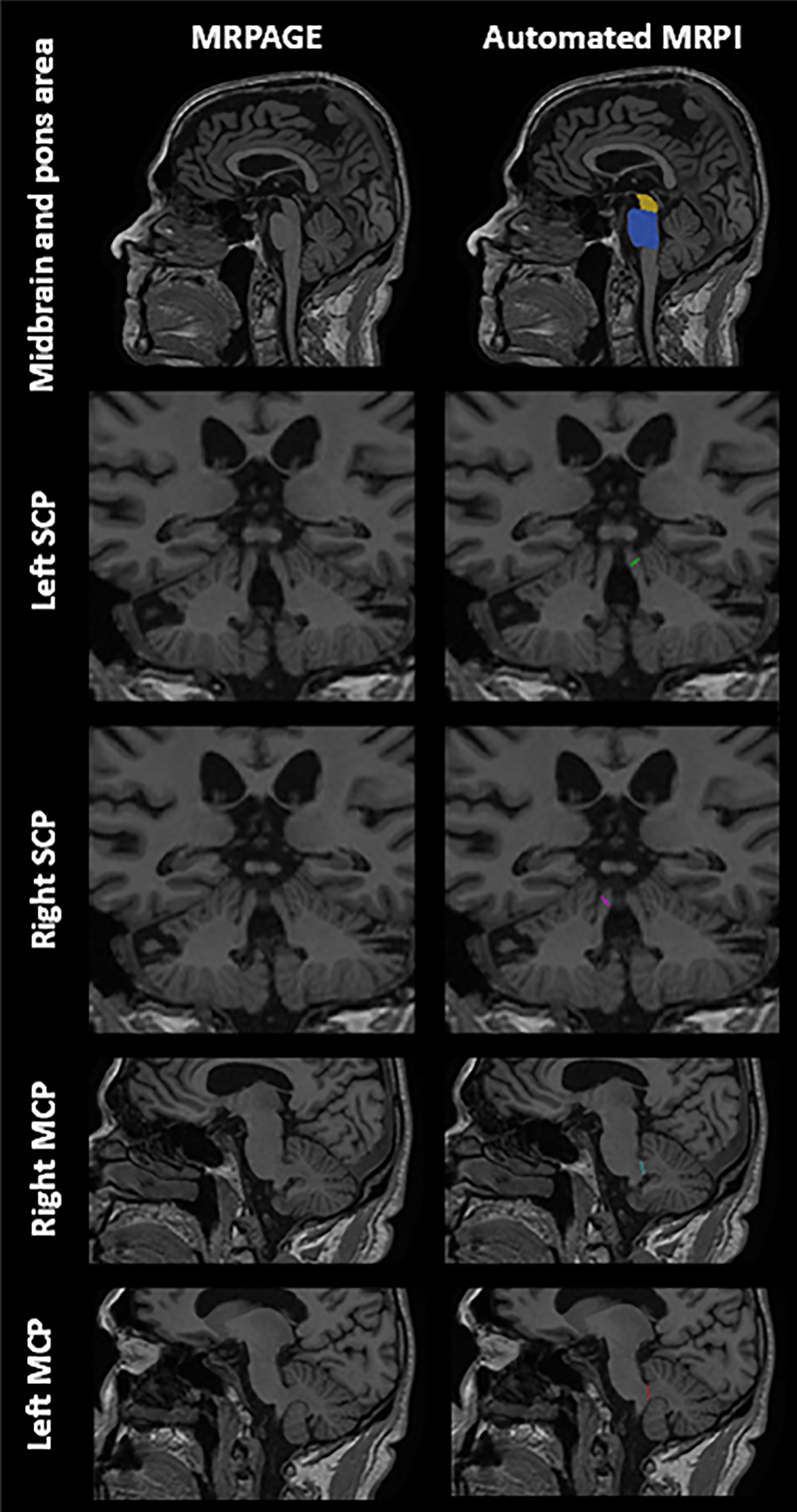
Fig. 1 Identical paired standard T1-weighted
images of a healthy 84 year-old male control illustrating automated anatomical placement
of color-coded landmarks for midbrain (yellow) and pons (blue) areas (first row),
followed by left and right SCP and MCP widths respectively. Abbreviation: MCP =
Middle Cerebellar Peduncle, SCP = Superior Cerebellar Peduncle.
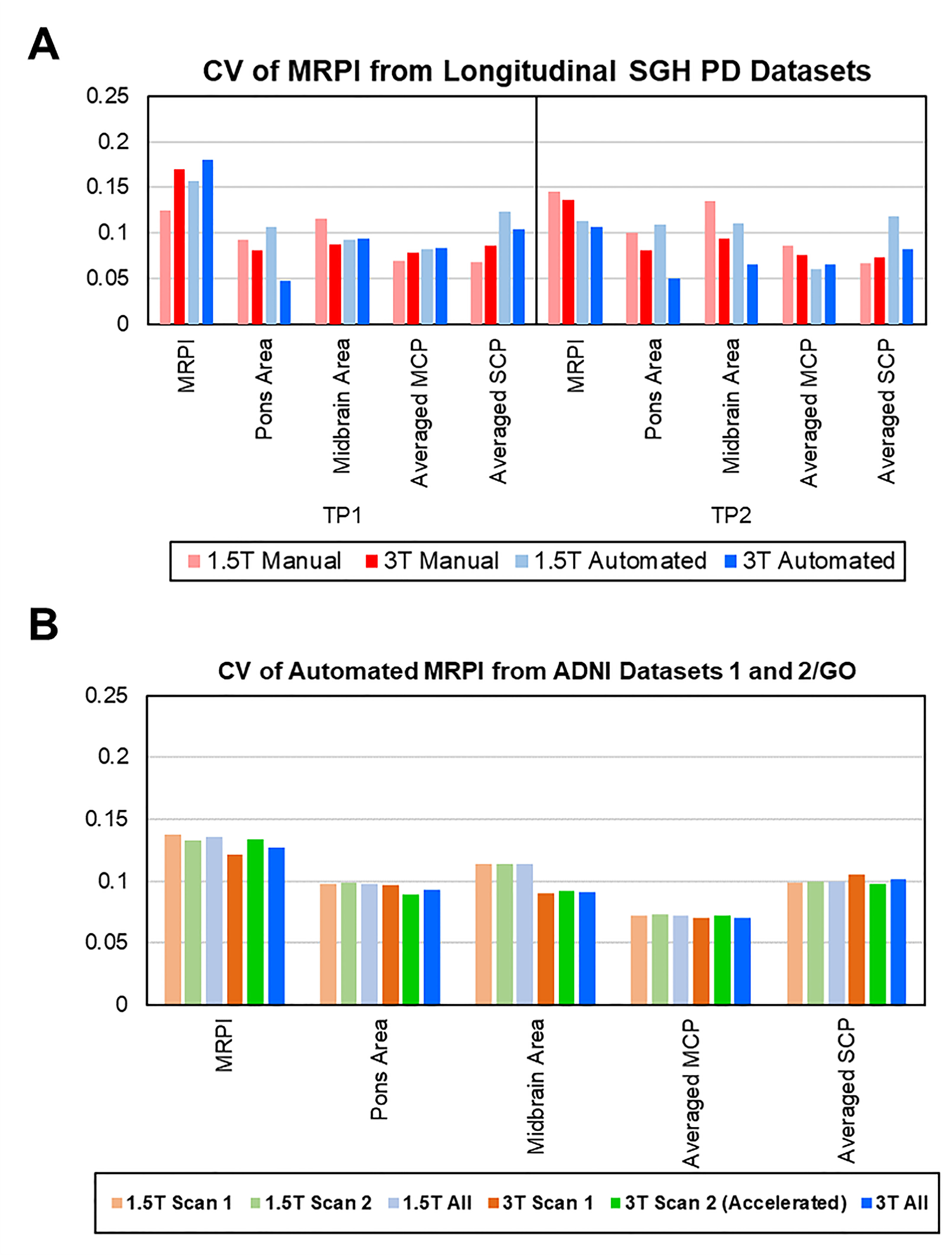
Fig. 3 Bar
charts of coefficients of variance (CV, y-axes) of values and subcomponents of
MRPI (x-axes) from (A) manual and automated MRPI derived from longitudinal SGH-PD
cohorts, and (B) automated MRPI derived from single TP ADNI1 (1.5T) and ADNI
2/GO (3T) cohorts. Abbreviation: MCP = Middle Cerebellar Peduncle, SCP =
Superior Cerebellar Peduncle, TP = Time Point.