Dmytro Pylypenko1, Yuhui Xiong1, Lanxin Ji1, Le He1, Yu Ma2, and Hua Guo1
1Center for Biomedical Imaging Research, Department of Biomedical Engineering, Tsinghua University, Beijing, China, 2Department of Neurosurgery, Tsinghua University Yuquan Hospital, Beijing, China
1Center for Biomedical Imaging Research, Department of Biomedical Engineering, Tsinghua University, Beijing, China, 2Department of Neurosurgery, Tsinghua University Yuquan Hospital, Beijing, China
In this study, we performed a comparative study of the vascular geometry in PD patients vs. AMC to quantify the structural changes in the cerebral vasculature by analyzing SWI MRA data.
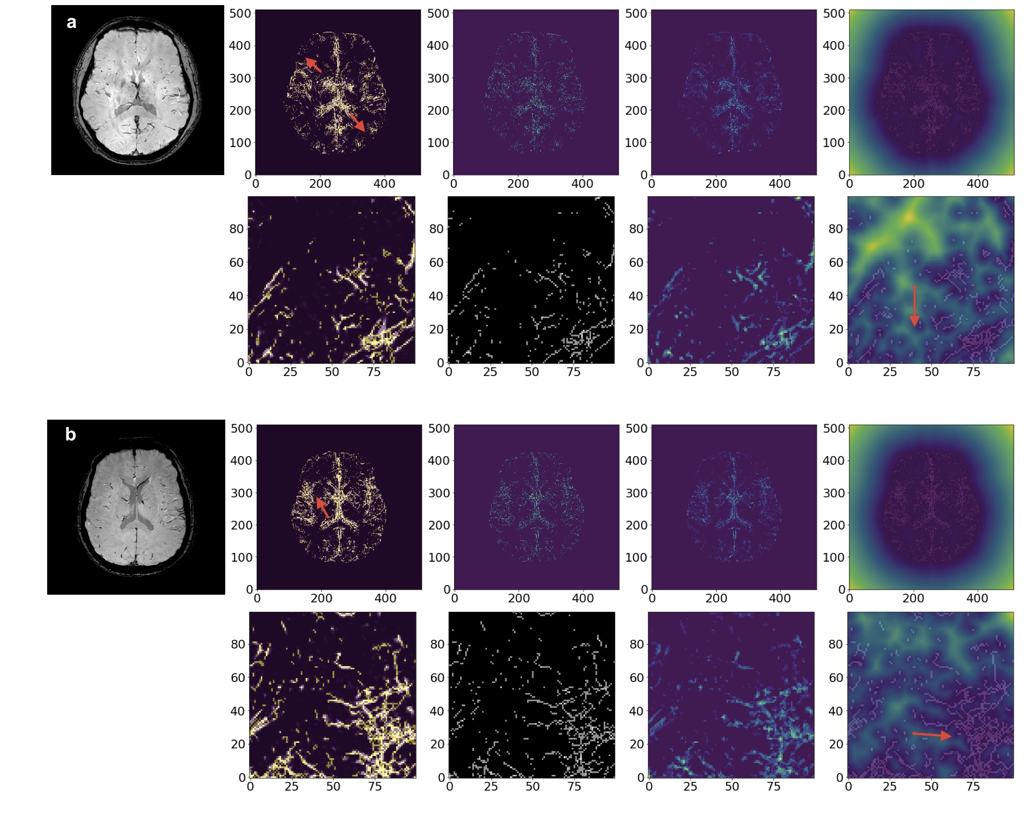
Figure 1. The results of the Segmentation and Skeletonization for the PD patients. a) Male PD patient, 63 years old. The venous tree is visually incomplete, the superficial cortical veins are missing (red arrowheads); b) Male PD patient, 74 years old. Increased tortuosity and fractal dimension (red arrowheads) of the venous system.

Figure 2. The results of the Segmentation and Skeletonization of the AMC, Female, 48 years old.