Alexandru V Avram1,2, Joelle E Sarlls3, and Peter J Basser1
1Eunice Kennedy Shriver National Institute of Child Health and Human Development, National Institutes of Health, Bethesda, MD, United States, 2Center for Neuroscience and Regenerative Medicine, The Henry Jackson Foundation, Bethesda,, MD, United States, 3National Institute of Neurological Disorders and Stroke, National Institutes of Health, Bethesda, MD, United States
1Eunice Kennedy Shriver National Institute of Child Health and Human Development, National Institutes of Health, Bethesda, MD, United States, 2Center for Neuroscience and Regenerative Medicine, The Henry Jackson Foundation, Bethesda,, MD, United States, 3National Institute of Neurological Disorders and Stroke, National Institutes of Health, Bethesda, MD, United States
We develop and test a clinical sequence with integrated IR
and isotropic diffusion encoding (IDE) preparations. T1-mean diffusivity (MD) correlation
spectra derived from whole-brain MRIs with a wide range of joint T1-MD weightings
show tissue-specific components healthy volunteers.
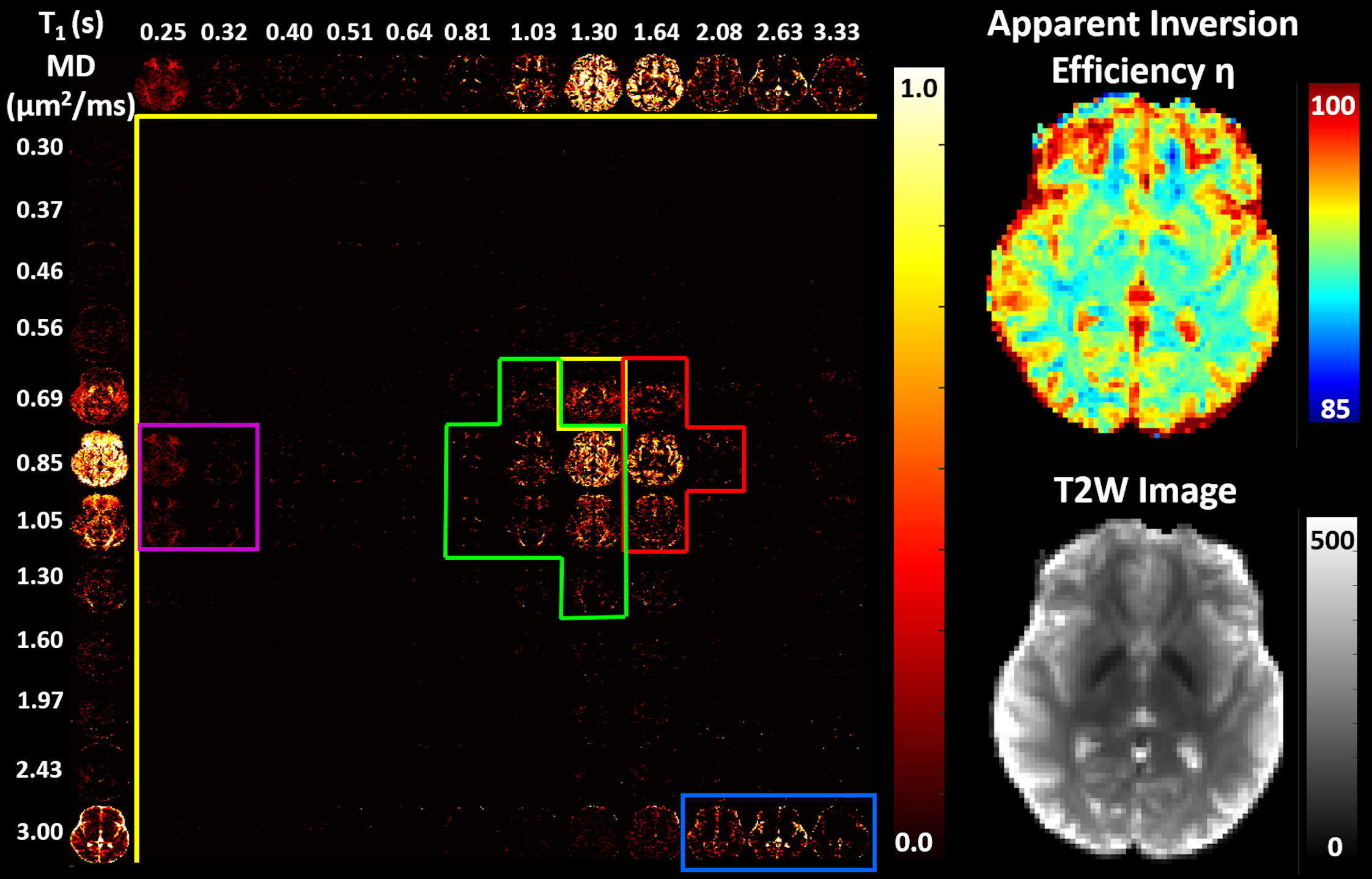
Figure 4: Left: Maps of 2D normalized
spectra of subvoxel T1-MD values along with the corresponding marginal
probability density functions (i.e., 1D normalized spectra) of subvoxel T1 values
(top row) and subvoxel MD values (left column) in a healthy volunteer. Spectral components specific to WM
(green), GM (red), subcortical GM (yellow), myelinated WM fibers (magenta), and
CSF (blue) can be observed both on the T1-D spectra as well as on the
corresponding marginal distributions. Right: Corresponding maps of the estimated apparent inversion efficiency η and non-attenuated signal.
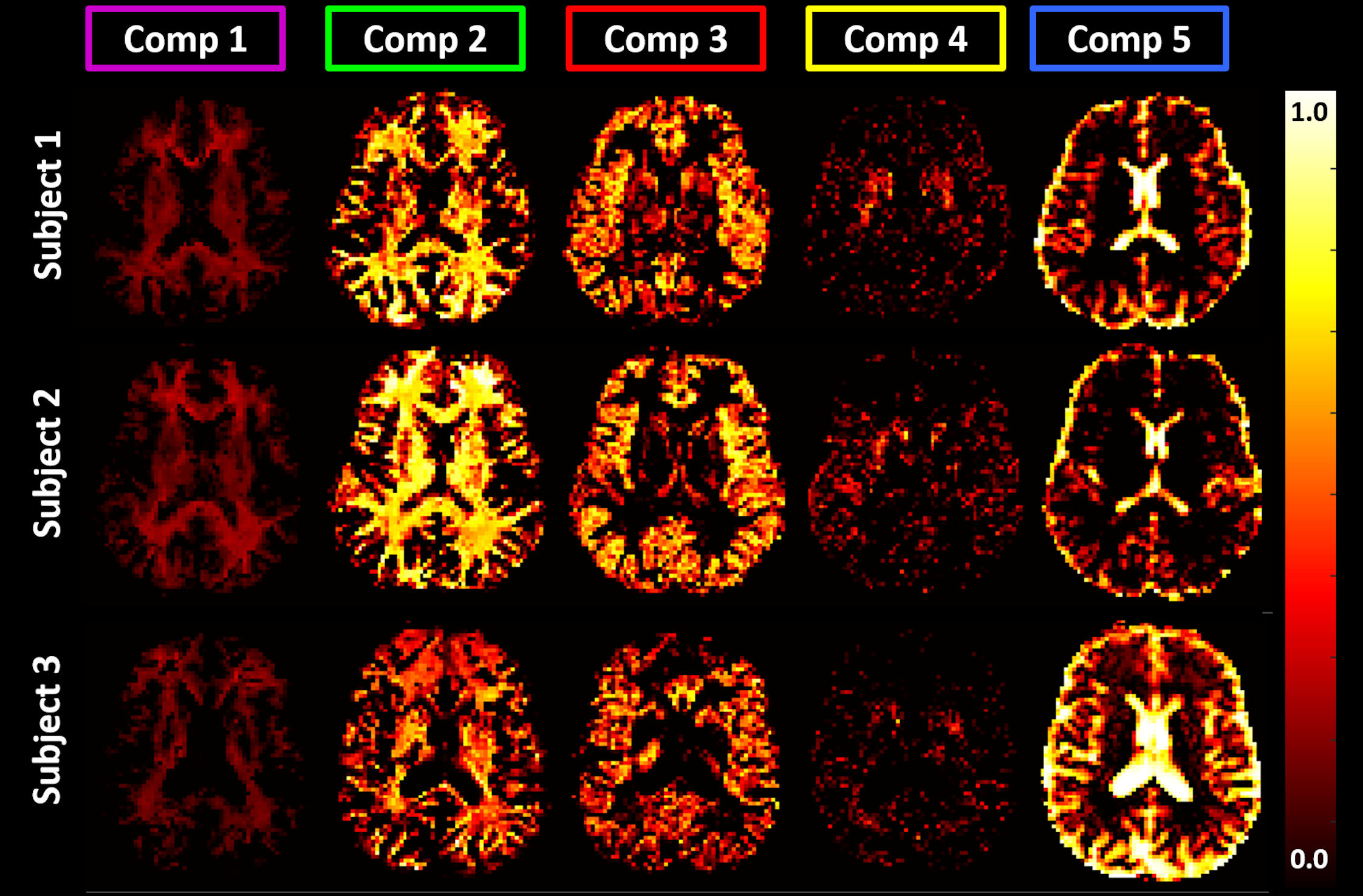
Figure 5: Maps of signal fractions corresponding to the principal
T1-MD spectral components delineated with color-coded boundaries in Fig. 4: Short-T1 WM (component 1) - magenta; WM (component 2) - green; GM (component
3) - red; Basal Ganglia (component 4) - yellow; and CSF (component 5) - blue.
Matching axial slices in three healthy volunteers show similar anatomical
features corresponding to these spectral domains.