Daiki Tamada1 and Scott B. Reeder1,2,3,4,5
1Radiology, University of Wisconsin-Madison, Madison, WI, United States, 2Medical Physics, University of Wisconsin-Madison, Madison, WI, United States, 3Biomedical Engineering, University of Wisconsin-Madison, Madison, WI, United States, 4Medicine, University of Wisconsin-Madison, Madison, WI, United States, 5Emergency Medicine, University of Wisconsin-Madison, Madison, WI, United States
1Radiology, University of Wisconsin-Madison, Madison, WI, United States, 2Medical Physics, University of Wisconsin-Madison, Madison, WI, United States, 3Biomedical Engineering, University of Wisconsin-Madison, Madison, WI, United States, 4Medicine, University of Wisconsin-Madison, Madison, WI, United States, 5Emergency Medicine, University of Wisconsin-Madison, Madison, WI, United States
A new phase-based T2 mapping method using a multi-echo
DESS with RF phase-modulation sequence was proposed. T2 can be estimated from the phase difference
of the acquired two
acquired echoes using the lookup table
approach that describes the relationship between T2 and signal phase.
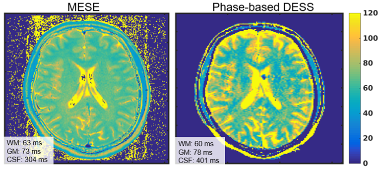
Figure
4: T2 map for a
volunteer brain measured using MESE and the proposed method. T2 values measured
using the proposed method for white matter (white dashed ROI, T2 = 60±11), grey matter (red dashed
ROI, T2 = 78±9.7), and
CSF (black dashed ROI, T2 = 401±186)
were close to those measured using MESE (T2 = 62±2.5, 73±2.8, and 354±70). However, artifacts
were observed in the lateral ventricle in the proposed method. Since the
sequence uses an unbalanced gradient moment, CSF pulsation may cause the artifacts.

Figure
1: Pulse sequence
diagram used in this study. Three-echo DESS sequence acquires FISP (S+1 and S+2) and PSIF (S1-) echoes. The two FISP echoes with different TEs enables
B0 map estimation which is used to demodulate B0 phase components of S+1 and S-1. RF
excitation is performed with a quadratic increase of transmitting phase to
encode T2 information into the phase of the signal. Calibration acquisition
using positive and negative readout gradient without phase-encoding is
incorporated to remove eddy current-induced phase error.