Dandan Peng1, Zhongshuai Zhang2, Cong Xia1, Yuancheng Wang1, and Shenghong Ju1
1Zhongda Hospital, Medical School of Southeast University, Nanjing, China, 2SIEMENS Healcare, Shanghai, China
1Zhongda Hospital, Medical School of Southeast University, Nanjing, China, 2SIEMENS Healcare, Shanghai, China
This study aimed to provide information
about differentiation of benign from malignant pulmonary lesions with IVIM using individual shimming technique among
33 lung lesions. The results initially indicated that ADC and IVIM and could be
great techniques for lung lesions detection.
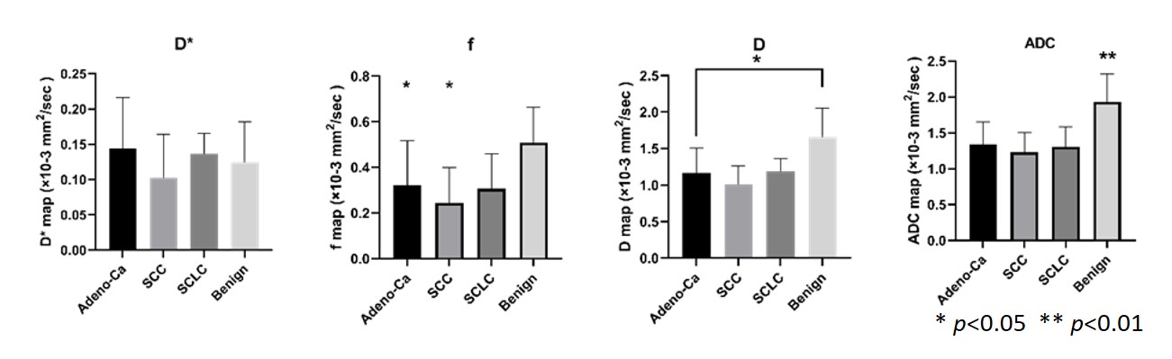
Figure 1. The IVIM quantitative values distribution of malignancy and benignity
in lung lesions. The mean ADC value of benign lesions is 1.846±0.403×10⁻³
mm2/s (ANOVA, p<0.01)
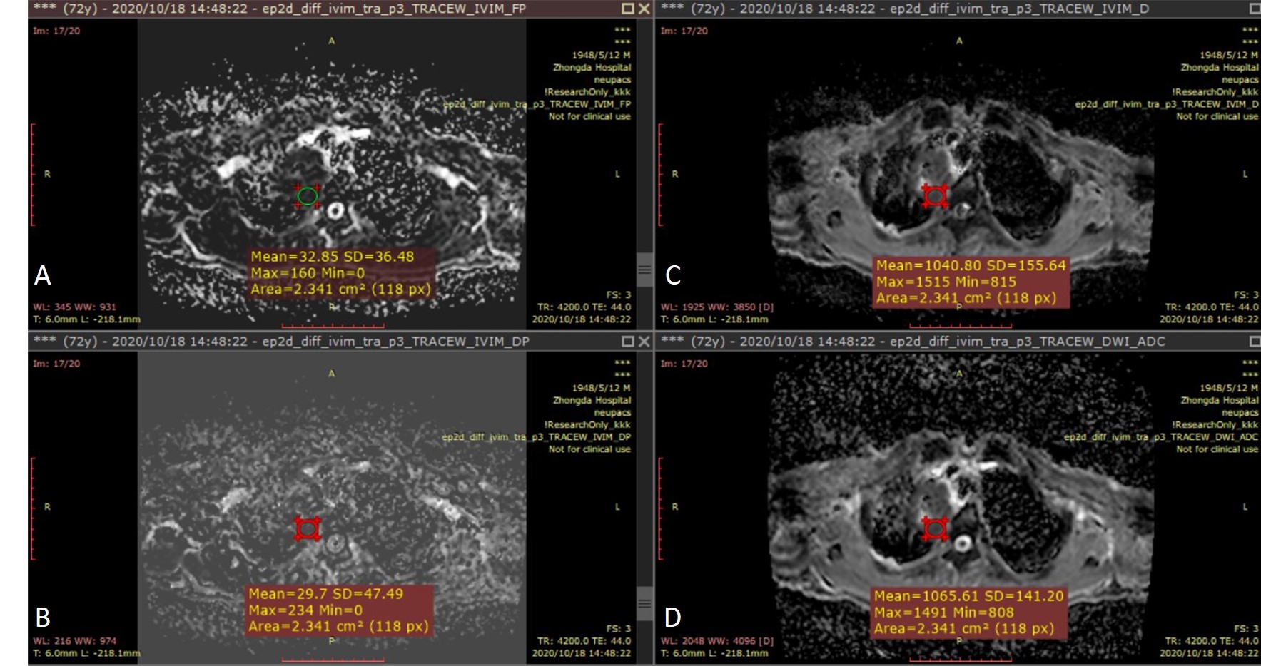
Figure 3. 72-year-old
male with a 6cm mass in the right upper lobe. This mass was diagnosed as
adenocarcinoma IIIb (cT4N2M0).ROI area of the mass is 2.341 cm2.(A):
IVIM-f shows a value of 0.033×10⁻³mm2/s
around the mass. (B): IVIM-D*
shows a value of 0.030×10⁻³mm2/s around
the mass. (C):IVIM-D shows a value of 1.041×10⁻³mm2/s around the mass. (D): ADC shows a value of 1.066×10⁻³mm2/s around the mass.