Ya-Jun Ma1, Michael Carl2, Hyungseok Jang1, Saeed Jerban1, Eric Y Chang1,3, Seth Kligerman1, and Jiang Du1
1UC San Diego, San Diego, CA, United States, 2GE Healthcare, San Diego, CA, United States, 3VA Health system, San Diego, CA, United States
1UC San Diego, San Diego, CA, United States, 2GE Healthcare, San Diego, CA, United States, 3VA Health system, San Diego, CA, United States
The 3D UTE Cones sequence with a randomized
ordering scheme can provide self-navigated and phase-resolved high-resolution
lung imaging, which may be potentially useful in clinical practice.
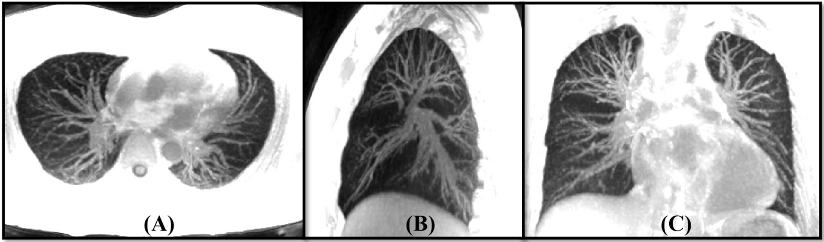
Figure 5. The
example of MIP lung images reconstructed from the end expiration phase in axial
(A), sagittal (B) and coronal (C) planes. The small pulmonary vessels are
well-displayed in these images.
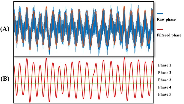
Figure 3. The
estimation of respiration using the phase of DC. The data were acquired from a
37-year-old healthy male volunteer. Panel A shows the raw phase of DC (i.e.,
the blue curve) received from a coil close to the liver. The red curve in panel
A is the corresponding low-pass filtered phase representing the respiration.
The cut-off frequency is ranged from 0.1 to 0.5 Hz. The respiration is segmented
into five phases (separated by the green lines) as shown in panel B. Phase 1 and 5 are corresponding to
the end of inspiration and expiration respectively.