Quan Dou1, Xue Feng1, Sohil Patel2, and Craig H. Meyer1
1Biomedical Engineering, University of Virginia, Charlottesville, VA, United States, 2Radiology & Medical Imaging, University of Virginia, Charlottesville, VA, United States
1Biomedical Engineering, University of Virginia, Charlottesville, VA, United States, 2Radiology & Medical Imaging, University of Virginia, Charlottesville, VA, United States
In this study, we analyzed the relationships between glioblastoma patients overall survival and several automatic segmentation-based MR imaging features. Results showed that combining imaging features with clinical factors improved the survival prediction.
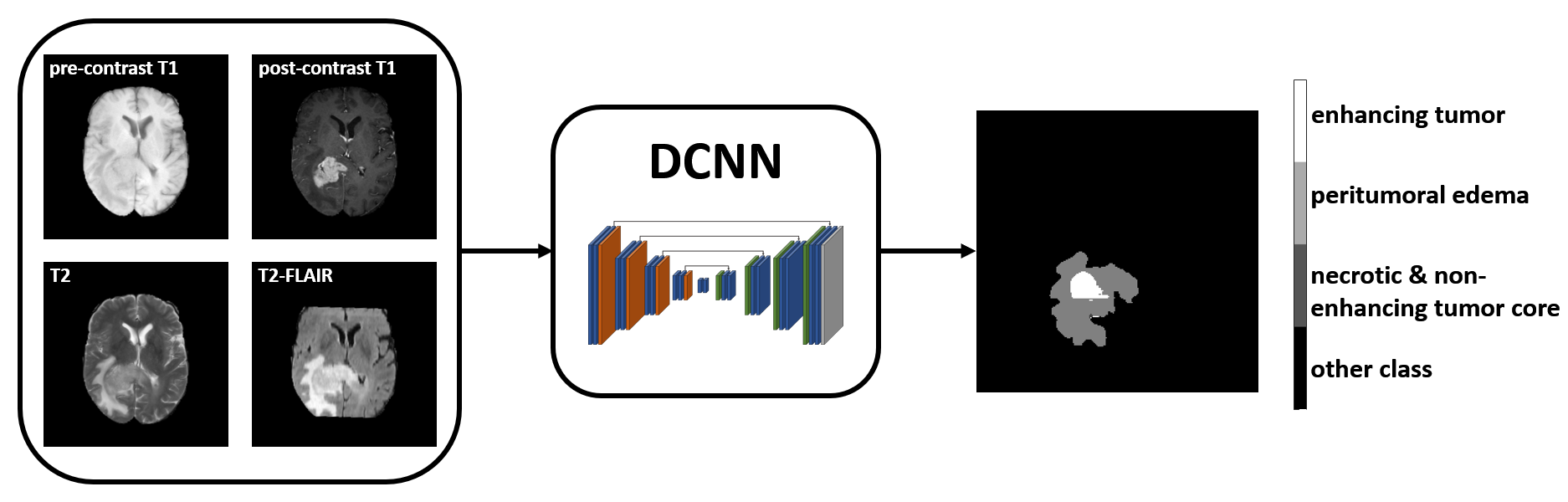
Figure 1. Deep learning-based automatic
segmentation. A pre-trained DCNN5 takes in pre- and post-contrast
T1-weighted, T2-weighted and T2-FLAIR images, and generates segmentation
results including three subregions: peritumoral edema, enhancing tumor, and
necrotic & non-enhancing tumor core.

Figure 3. Receiver operating characteristic
curve analyses for OS classification models.