Atsuro Suzuki1, Chizue Ishihara1, Yukio Kaneko1, Tomoki Amemiya1, Yoshitaka Bito1, and Toru Shirai1
1Healthcare Business Unit, Hitachi, Ltd., Kokubunji-shi, Japan
1Healthcare Business Unit, Hitachi, Ltd., Kokubunji-shi, Japan
To reduce
the inhomogeneous noise, we developed a noise reduction method by using
multiple convolutional
neural networks (CNNs) optimized for noise intensity. Denoised brain
images demonstrated improved mean square error (MSE) and signal to noise ratio (SNR) throughout the
brain regions.

Deep-learning-based
noise reduction incorporating spatial distribution of noise. Parallel imaging creates
a low sensitivity region in the center of the reconstructed image (indicated by
the dashed line in the g-factor map), so the thresholds segmenting the high
g-factor region in the g-factor map include the central low sensitivity region.
The two g-factor regions were segmented by using thresholds of 1.4 and 2.0 for
R of 3 and 4, respectively.
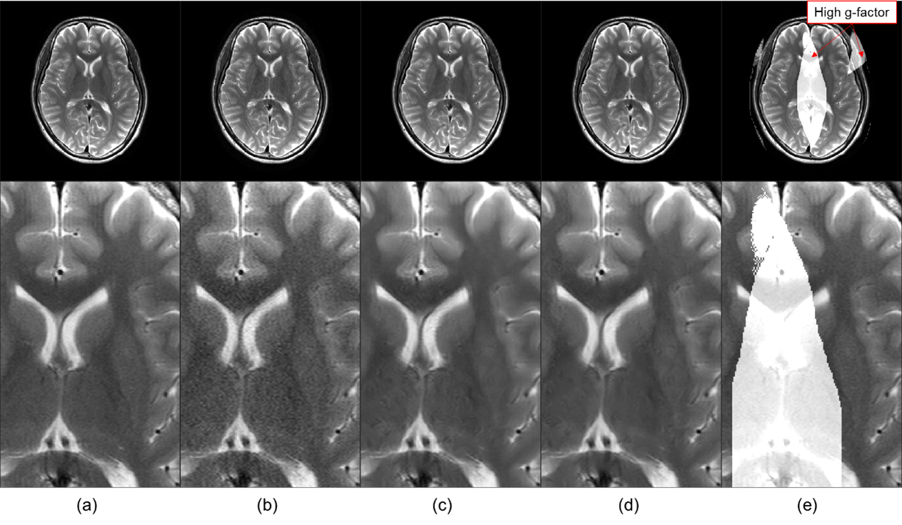
T2
weighed brain image: (a) full sampling image, (b) input image with acceleration
rate of 3; input image denoised by (c) conventional method and (d) MA-CNNR; (e)
high g-factor region registered in full sampling image. Due to the higher
g-factor, the noise in the central region of the input image (b) was higher
than that in the peripheral region.