Rajiv S Deshpande1,2, Michael C Langham2, and Felix W Wehrli2
1Dept. of Bioengineering, University of Pennsylvania, Philadelphia, PA, United States, 2Dept. of Radiology, University of Pennsylvania, Philadelphia, PA, United States
1Dept. of Bioengineering, University of Pennsylvania, Philadelphia, PA, United States, 2Dept. of Radiology, University of Pennsylvania, Philadelphia, PA, United States
A T2-based oximetry technique is presented that quantifies whole-organ
metabolic rate of oxygen by simultaneously measuring blood flow velocity and T2
of blood water protons at a single anatomic location in 18 seconds scan time.
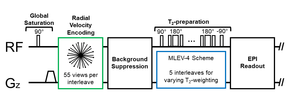
Figure 1: Pulse sequence timeline showing interleaved radial phase-contrast MRI preceding
a background-suppressed T2-prepared EPI readout to simultaneously
measure blood flow velocity and T2 (and SvO2),
respectively. 55 radial views are acquired in each of the five interleaves (each
corresponding to a TE).
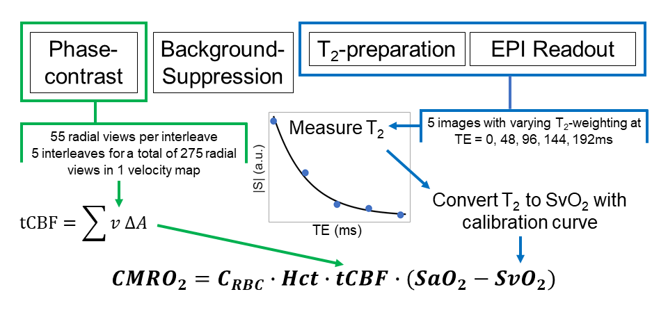
Figure 2: Pulse sequence workflow: phase-contrast
consists of 55 radial views in each interleave. There are five interleaves for
275 total views in one velocity map to determine total cerebral blood flow (tCBF).
Each interleave generates an image with a different T2-weighting,
from which T2 is measured and then converted to SvO2.