Paul Han1, Thibault Marin1, Vanessa Landes2, Yanis Djebra1,3, Georges El Fakhri1, and Chao Ma1
1Gordon Center for Medical Imaging, Department of Radiology, Massachusetts General Hospital and Harvard Medical School, Boston, MA, United States, 2GE Healthcare, Boston, MA, United States, 3LTCI, Télécom Paris, Institut Polytechnique de Paris, Paris, France
1Gordon Center for Medical Imaging, Department of Radiology, Massachusetts General Hospital and Harvard Medical School, Boston, MA, United States, 2GE Healthcare, Boston, MA, United States, 3LTCI, Télécom Paris, Institut Polytechnique de Paris, Paris, France
This work investigated effects of B1 inhomogeneity in the context of T1 estimation, considering B1 inhomogeneity in the model for T1 estimation, and characterized effects of flip angle and heart rate to optimize a spoiled GRE-based ECG-gated IR acquisition scheme for 3D cardiac T1 mapping.

Figure 4. Phantom
study results. A:
Estimated T1 maps from IR-FSE,
MOLLI, and ECG-gated IR schemes of 8-8, 5-(3)-5-(3), and 10-(3)-10-(3) with
spoiled GRE. B: Scatter plots of T1 from MOLLI and ECG-gated IR schemes
of 8-8, 5-(3)-5-(3), and 10-(3)-10-(3) with spoiled GRE. Solid line represents
line of identity and dashed line represents line of regression. C:
Bland-Altman plots from MOLLI and 8-8, 5-(3)-5-(3), and 10-(3)-10-(3) schemes
with spoiled GRE. Solid line represents mean difference and dashed line
represents the 95% confidence interval for limits of agreement.
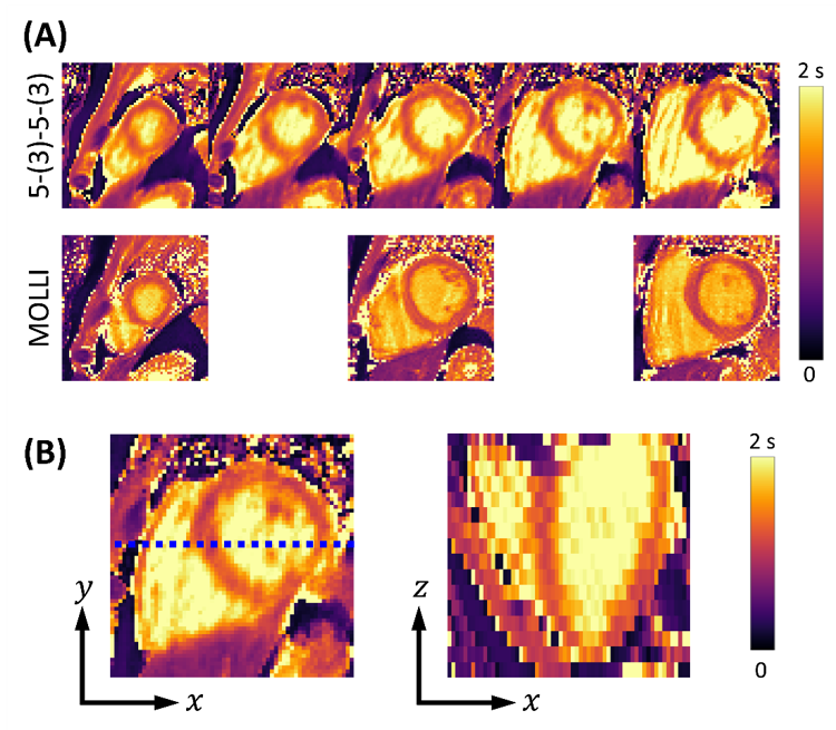
Figure 5. In
vivo study results. A:
3D T1 maps from 5-(3)-5-(3) scheme with spoiled GRE and 2D T1
maps from MOLLI. B: Slice view of the 3D T1 map from 5-(3)-5-(3)
scheme with spoiled GRE along the blue dotted line. The estimated T1
in the apical, mid-cavity, and basal regions of the myocardium were 1165.9 ±
109.4, 1166.1 ± 85.5, and 1256.8 ± 162.6 ms from 5-(3)-5-(3) scheme with
spoiled GRE and 1195.6 ± 118.7, 1185.8 ± 116.7, and 1115.0 ± 135.6 ms from
MOLLI, respectively.