Laura Saunders1, Paul J. C. Hughes1, James Eaden1, Andy J Swift1, Stephen Bianchi2, and Jim M Wild1
1Infection Immunity and Cardiovascular Disease, University of Sheffield, Sheffield, United Kingdom, 2Sheffield Teaching Hospitals NHS, Sheffield, United Kingdom
1Infection Immunity and Cardiovascular Disease, University of Sheffield, Sheffield, United Kingdom, 2Sheffield Teaching Hospitals NHS, Sheffield, United Kingdom
A comparison Look-Locker and variable flip angle T1 mapping sequences in phantoms
and in vivo, in the lung, liver and blood. In vivo, there are
significant differences in measured T1 between inversion recovery
and VFA sequences, which differ in different tissues.
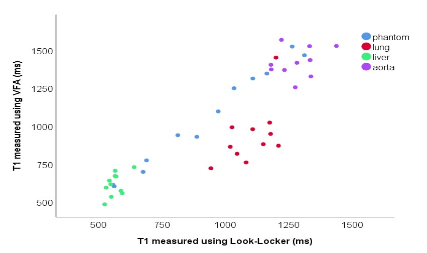
Figure
3: Scatter plot showing the correlation in lung T1 measured using the VFA
acquisition and Look-Locker acquisition, for both phantom and in vivo data. Regions of interest were
drawn in the lung, liver and descending aorta (blood) for each participant. 4
participants were healthy volunteers, 7 were patients with IPF.
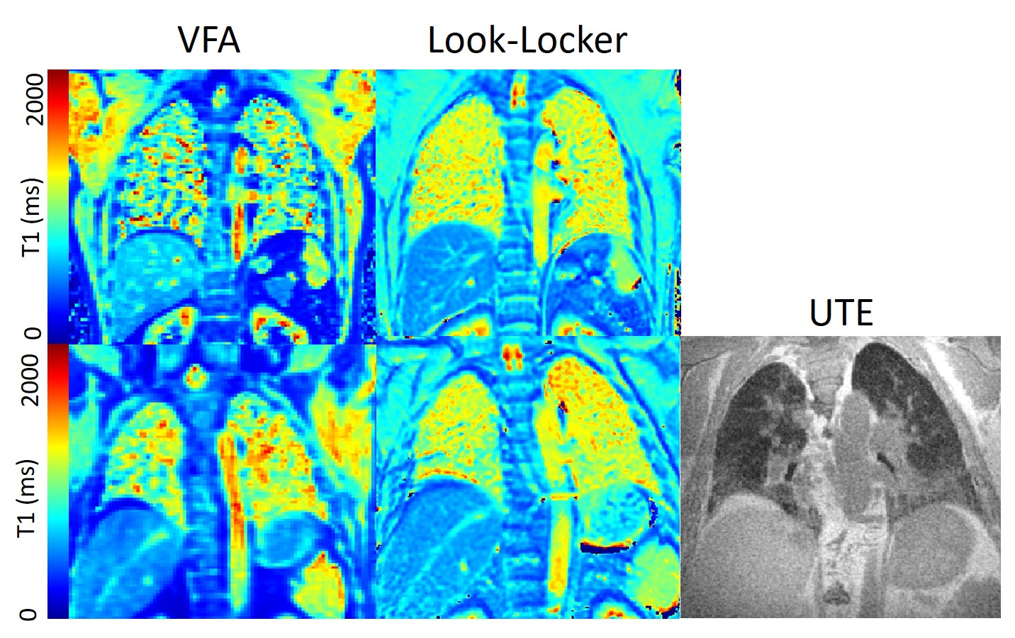
Figure
2: Example T1 maps in a healthy volunteer and patient with IPF using
Look-Locker and VFA acquisitions. A similar-slice UTE image is also shown for
the patient, to visualise regions of increases lung density.