YANLI JIANG1, Pin Yang1, FengXian Fan1, Wanjun Hu1, Jing Zhang1, and Shaoyu Wang2
1Department of Magnetic Resonance, LanZhou University Second Hospital, LanZhou, China, 2MR Scientific Marketing, Siemens Healthineers, Shanghai, China
1Department of Magnetic Resonance, LanZhou University Second Hospital, LanZhou, China, 2MR Scientific Marketing, Siemens Healthineers, Shanghai, China
Our study used B1 corrected native T1 value to
assess their diagnostic accuracy for staging of liver fibrosis. The results
show that native T1 values had potential value for staging of liver fibrosis.

Figure 2, Scatter plots show a significantly
positive correlation between Fibroscan and T1 values (r=0.398,
P=0.04)
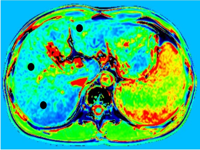
Figure 1, Examples of region of interest (ROI) placement in a patient graded as G1S1. Three ROIs were placed in the different area of liver to measure the T1 relaxation time(● represent the ROI )