Nathan Tibbitts Roberts1,2, Ruvini Navaratna1,3, Diego Hernando1,3, and Scott B Reeder1,3,4,5,6
1Radiology, University of Wisconsin - Madison, Madison, WI, United States, 2Electrical and Computer Engineering, University of Wisconsin - Madison, Madison, WI, United States, 3Medical Physics, University of Wisconsin - Madison, Madison, WI, United States, 4Biomedical Engineering, University of Wisconsin - Madison, Madison, WI, United States, 5Medicine, University of Wisconsin - Madison, Madison, WI, United States, 6Emergency Medicine, University of Wisconsin - Madison, Madison, WI, United States
1Radiology, University of Wisconsin - Madison, Madison, WI, United States, 2Electrical and Computer Engineering, University of Wisconsin - Madison, Madison, WI, United States, 3Medical Physics, University of Wisconsin - Madison, Madison, WI, United States, 4Biomedical Engineering, University of Wisconsin - Madison, Madison, WI, United States, 5Medicine, University of Wisconsin - Madison, Madison, WI, United States, 6Emergency Medicine, University of Wisconsin - Madison, Madison, WI, United States
This work demonstrates the combination of simultaneous multislice (SMS) imaging to accelerate 2D motion robust CSE-MRI acquisitions. Results confirm the feasibility of combining these two techniques to achieve rapid, whole-liver, motion robust quantitative tissue characterization.
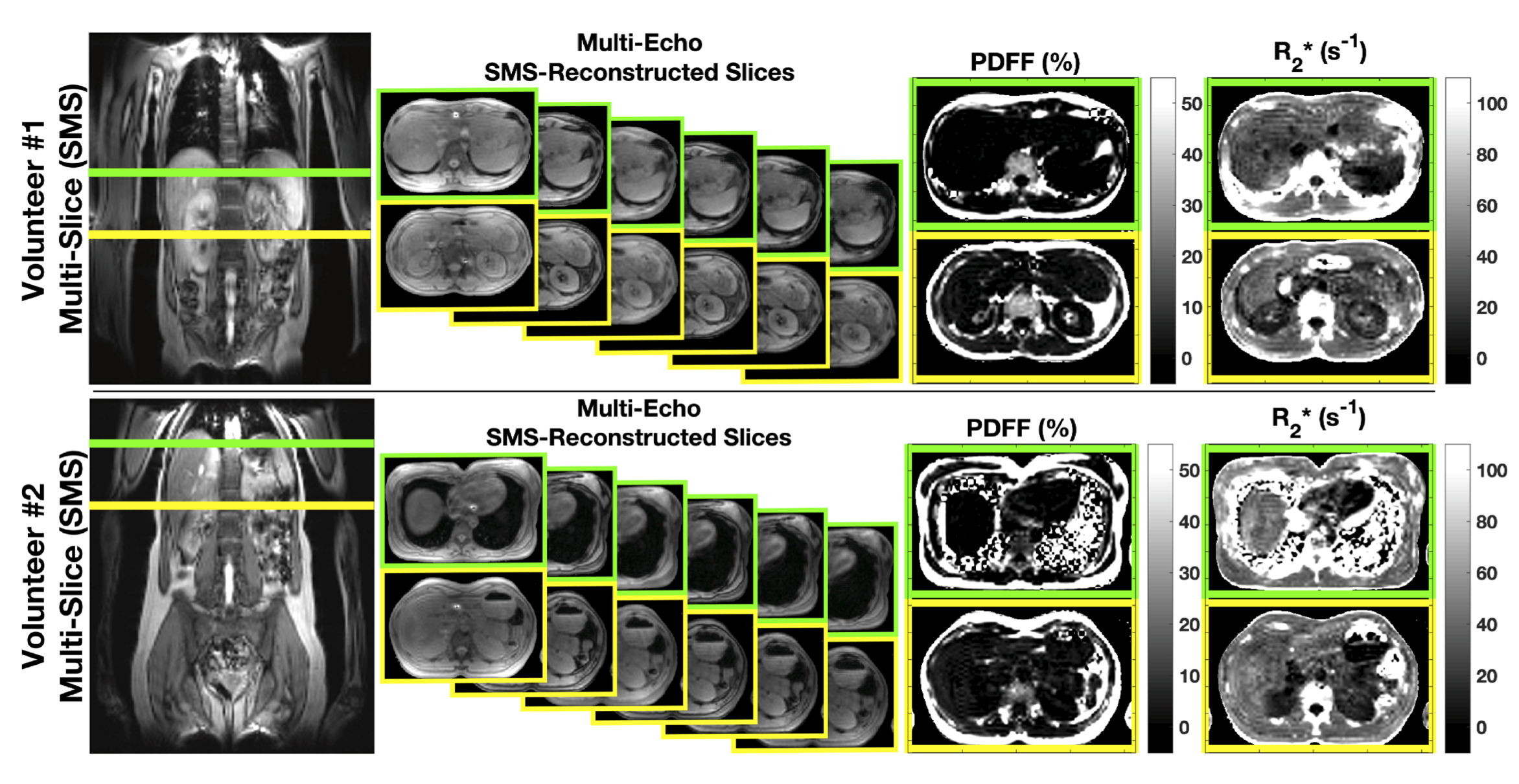
Figure 3. Results from two healthy volunteers demonstrate the feasibility of combining SMS with CSE-MRI to achieve rapid, whole-liver, motion robust tissue quantification. The far-left image shows the physical locations of the simultaneously excited slices. The middle set of images shows the multi-echo separated images, followed by the estimated PDFF and R2* maps for both slices.
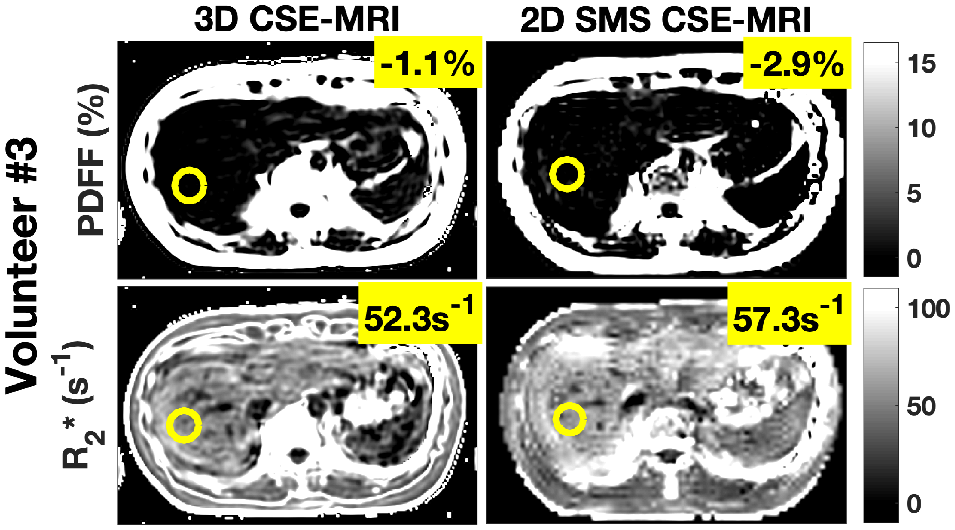
Figure 4. Comparison to a reference 3D CSE-MRI shows good agreement with the 2D SMS CSE-MRI protocol for estimated PDFF and R2*. Averages from each region of interest are shown. Note the negative values in the PDFF maps can likely be attributed to noise related bias common in low SNR CSE-MRI PDFF maps estimated with a fully complex signal model (see reference 6).